LITHUANIAN UNIVERSITY OF HEALTH SCIENCES VETERINARY ACADEMY. Jūratė Stankevičienė
|
|
|
- Medėja Gailius
- prieš 4 metus
- Peržiūrų:
Transkriptas
1 LITHUANIAN UNIVERSITY OF HEALTH SCIENCES VETERINARY ACADEMY Jūratė Stankevičienė CHARACTERISTICS AND RESISTANCE TO ANTIMICROBIALS OF STAPHYLOCOCCUS AUREUS AND STAPHYLOCOCCUS PSEUDINTERMEDIUS ISOLATED FROM PET ANIMALS AND THEIR OWNERS Doctoral Dissertation Agricultural Sciences, Veterinary (A 002) Kaunas, 2019
2 Dissertation has been prepared at the Department of Veterinary Pathobiology (former Infectious Diseases) of the Veterinary Academy of the Lithuanian University of Health Sciences during the period of Scientific Supervisor Prof. Dr. Jūratė Šiugždaitė (Lithuanian University of Health Sciences, Agricultural Sciences, Veterinary A 002). Dissertation is defended at the Veterinary Research Council of the Lithuanian University of Health Sciences: Chairperson Prof. Dr. Alius Pockevičius (Lithuanian University of Health Sciences, Agricultural Sciences, Veterinary A 002). Members: Assoc. Prof. Dr. Gintaras Daunoras (Lithuanian University of Health Sciences, Agricultural Sciences, Veterinary A 002); Dr. Marius Virgailis (Lithuanian University of Health Sciences, Agricultural Sciences, Veterinary A 002); Prof. Dr. Algimantas Paulauskas (Vytautas Magnus University, Natural Sciences, Biology N 010); Dr. Olga Muter (University of Latvia, Natural Sciences, Biology N 010). Dissertation will be defended at the open session of the Veterinary Research Council of Lithuanian University of Health Sciences, Veterinary Academy, 2019 on 30 of August at 10 p.m. in the Dr. S. Jankauskas auditorium. Address: Tilžės 18, LT-47181, Kaunas, Lithuania.
3 LIETUVOS SVEIKATOS MOKSLŲ UNIVERSITETAS VETERINARIJOS AKADEMIJA Jūratė Stankevičienė STAPHYLOCOCCUS AUREUS IR STAPHYLOCOCCUS PSEUDINTERMEDIUS, IŠSKIRTŲ IŠ GYVŪNŲ AUGINTINIŲ IR JŲ SAVININKŲ, CHARAKTERIZAVIMAS IR ATSPARUMAS ANTIMIKROBINĖMS MEDŽIAGOMS Daktaro disertacija Žemės ūkio mokslai, veterinarija (A 002) Kaunas, 2019
4 Disertacija rengta metais Lietuvos sveikatos mokslų universitete, Veterinarijos akademijoje, Veterinarinės patobiologijos katedroje (buvusi Užkrečiamųjų ligų katedra), Mikrobiologijos laboratorijoje. Mokslinė vadovė prof. dr. Jūratė Šiugždaitė (Lietuvos sveikatos mokslų universitetas, že mės ūkio mokslai, veterinarija A 002). Disertacija ginama Lietuvos sveikatos mokslų universiteto Veterinarijos mokslo krypties taryboje: Pirmininkas prof. dr. Alius Pockevičius (Lietuvos sveikatos mokslų universitetas, žemės ūkio mokslai, veterinarija A 002). Nariai: doc. dr. Gintaras Daunoras (Lietuvos sveikatos mokslų universitetas, žemės ūkio mokslai, veterinarija A 002); dr. Marius Virgailis (Lietuvos sveikatos mokslų universitetas, žemės ūkio mokslai, veterinarija A 002); prof. dr. Algimantas Paulauskas (Vytauto Didžiojo universitetas, gamtos mokslai, biologija N 010); dr. Olga Muter (Latvijos universitetas, gamtos mokslai, biologija N 010). Disertacija ginama viešame Veterinarijos mokslo krypties tarybos posėdyje 2019 m. rugpjūčio 30 d. 10 val. Lietuvos sveikatos mokslų universiteto Veterinarijos akademijos dr. S. Jankausko auditorijoje. Disertacijos gynimo vietos adresas: Tilžės g. 18, LT-47181, Kaunas, Lietuva.
5 CONTENTS ABBREVIATIONS... 8 INTRODUCTION LITERATURE REVIEW History Taxonomy Colonization of Staphylococcus aureus and Staphylococcus pseudintermedius Staphylococcus aureus in canine and feline Staphylococcus aureus in human Staphylococcus pseudintermedius in dogs and cats Staphylococcus pseudintermedius in human Staphylococcus aureus and Staphylococcus pseudintermedius as pathogens Diseases caused by Staphylococcus aureus and Staphylococcus pseudintermedius in dogs and cats Diseases caused by Staphylococcus aureus and Staphylococcus pseudintermedius in human General characteristics and biochemical properties of Staphylococcus aureus and Staphylococcus pseudintermedius Virulence factors of staphylococci Identification of staphylococci Phenotypic identification Genotypic identification DNA sequencing Resistance to antimicrobials Antimicrobial resistance of Staphylococcus pseudintermedius Antimicrobial resistance of Staphylococcus aureus Antimicrobial susceptibility testing Interspecies transmission of staphylococci MATERIAL AND METHODS Collection of bacterial samples Isolation of pure culture of the staphylococci Phenotypic identification of staphylococci Detection of coagulase-positive staphylococci Determination of virulence factors for Staphylococcus aureus and Staphylococcus pseudintermedius
6 Biochemical characteristics of Staphylococcus aureus and Staphylococcus pseudintermedius Genotypic identification of Staphylococcus aureus and Staphylococcus pseudintermedius Antimicrobial susceptibility of Staphylococcus aureus and Staphylococcus pseudintermedius Antimicrobial resistance of Staphylococcus aureus and Staphylococcus pseudintermedius detection by disc diffusion method The minimal inhibitory and minimum bactericidal concentrations of antimicrobials against Staphylococcus aureus and Staphylococcus pseudintermedius Genotypic identification of resistance genes in Staphylococcus aureus and Staphylococcus pseudintermedius Statistical analysis Sequencing and phylogenetic analysis RESULTS Description of population Isolation of Staphylococcus aureus and Staphylococcus pseudintermedius Detection of virulence factors in Staphylococcus aureus and Staphylococcus pseudintermedius Biochemical identification of Staphylococcus aureus and Staphylococcus pseudintermedius Genotypic identification of Staphylococcus aureus and Staphylococcus pseudintermedius Prevalence of Staphylococcus aureus and Staphylococcus pseudintermedius Staphylococcus aureus in canine Staphylococcus pseudintermedius in canine Staphylococcus aureus in feline Staphylococcus pseudintermedius in feline Staphylococcus aureus in humans Staphylococcus pseudintermedius in humans Phylogenetic analysis of Staphylococcus aureus and Staphylococcus pseudintermedius Staphylococci genome comparisons Phenotypic antimicrobial susceptibility of Staphylococcus aureus and Staphylococcus pseudintermedius
7 Resistance of Staphylococcus pseudintermedius in companion animals Resistance of Staphylococcus pseudintermedius in humans Resistance of Staphylococcus aureus in companion animals Resistance of Staphylococcus aureus in humans The minimal inhibitory and minimal bactericidal concentrations of antimicrobials against Staphylococcus aureus and Staphylococcus pseudintermedius Genotypic identification of resistance genes in Staphylococcus aureus and Staphylococcus pseudintermedius DISCUSSION CONCLUSIONS RECOMMENDATIONS BIBLIOGRAPHY PUBLICATIONS SANTRAUKA ANNEXES CURRICULUM VITAE ACKNOWLEDGEMENTS
8 ABBREVIATIONS AML amoxicillin AMP ampicillin AUG Augmentin Bp base pair BPA Baird Parker agar C cytosine CFU/mL colony-forming units per milliliter CLSI The Clinical and Laboratory Standards Institute CNS coagulase-negative staphylococci CPS coagulase-positive staphylococci CI confidence interval DNA deoxyribonucleic acid EUCAST The European Committee on Antimicrobial Susceptibility Testing G guanine Luk leucocidin toxins M-PCR multiplex polymerase chain reaction MBC minimum bactericidal concentration MDR multidrug resistant MIC minimal inhibitory concentration mol% mole percent MRSA methicillin-resistant Staphylococcus aureus MRSP methicillin-resistant Staphylococcus pseudintermedius MSSA methicillin-sensitive Staphylococcus aureus ONPG ortho-nitrophenyl-galactopyranoside OR odds ratio OX oxacillin PBP penicillin binding protein PCR polymerase chain reaction PFGE pulsed field gel electrophoresis PG penicillin G PVL Panton Valentine leucocidin PYR L-pyrrolidonyl-β-naphtylamide rmp revolutions per minute RNA ribonucleic acid S. aureus Staphylococcus aureus S. delphini Staphylococcus delphini S. hyicus Staphylococcus hyicus S. intermedius Staphylococcus intermedius S. lutrae Staphylococcus lutrae S. pseudintermedius Staphylococcus pseudintermedius S. schleiferi subsp. coagulans Staphylococcus schleiferi subspecies coagulans SCCmec Staphylococcal Cassette Chomosome mec SE staphylococcal enterotoxins SIG Staphylococcus intermedius group spp species 8
9 INTRODUCTION Dogs and cats probably are the most popular pet animals in Europe. The relationship between companion animals and humans has changed through the years. While in the past pets usually were maintained outside households, today they are often kept inside houses. Close physical contact by touching, petting and licking occurs at high frequency on the basis of the current perception of household pets as actual family members. Companion animals and humans can act as reservoirs of resistant to many antimicrobials bacteria [71]. Staphylococci are Gram-positive spherical bacteria of enormous clinical and biotechnological relevance [94]. Staphylococcal infections are common to veterinary and human medicine. β-lactam antibiotics are among frequently prescribed antibiotics worldwide to treat staphylococcal infections. Antimicrobial resistance is changing over time and is generally rising steadily for those antimicrobials that are often used. According to the published academic writings, the transfer of Staphylococcus pseudintermedius between human and zoonotic hosts is possible. There are several reports of serious MRSP-infections in humans [3, 4]. The situation is even more complicated, because the precise identification of Staphylococcus pseudintermedius requires genetic methods that are rarely used in routine diagnosis [15, 108]. It should be noted that this specie of bacteria until the year 2005 was identified and called as Staphylococcus intermedius or even Staphylococcus aureus [106]. Consequently, the importance of this bacterium as a pathogen appears to be underestimated [124]. The aim of the study is: To determine the prevalence of Staphylococcus aureus and Staphylococcus pseudintermedius colonization in companion animals and humans and their resistance to antimicrobials. Objectives of the study: 1. To isolate and identify Staphylococcus aureus and Staphylococcus pseudintermedius in the populations of companion animals and their owners using genotypic techniques. 2. To evaluate risk factors for pets and their owners for becoming carriers of Staphylococcus aureus and Staphylococcus pseudintermedius. 3. To determine phylogenetic relationship between isolated Staphylococcus aureus and Staphylococcus pseudintermedius strains. 9
10 4. To evaluate antimicrobial resistance of Staphylococcus aureus and Staphylococcus pseudintermedius Practical significance and scientific novelty The study was conducted to determine the prevalence of pathogenic staphylococci in pet animals and their owners. While Staphylococcus pseudintermedius have been recently distinguished as a new pathogenic species, the data on methicillin-resistant strains and resistance to other antimicrobial agents of these bacteria in this country are insufficient. As empirical treatment is often applied in veterinary clinics, antimicrobials are used to prevent secondary infection; it is useful to know which antimicrobials work best against pathogenic staphylococci. It is popular to keep one or even a few pets in one household in this country. A close relationship is often observed between a pets and pet owners. It is necessary to investigate the factors that influence the transmission of pathogenic bacteria between pets and their breeders in order to prevent the transmission of pathogenic and resistant bacteria between each other. Colonization of Staphylococcus pseudintermedius strains was detected not only in pets but also in humans. It is considered as a zoonosis. Staphylococcus aureus and Staphylococcus pseudintermedius strains containing the blaz gene which encodes resistance to β-lactam antibiotics were identified during the study. Phylogenetic analysis of Staphylococcus aureus and Staphylococcus pseudintermedius isolated from humans and their pets living in the same household was carried out in this study. 10
11 1. LITERATURE REVIEW 1.1. History The importance of staphylococci as pathogens for human and animals has been recognized for more than 100 years. The name Staphylococcus (staphyle, bunch of grapes) was introduced by Ogston (1883) for the group micrococci causing inflammation and suppuration. He was the first to differentiate two kinds of pyogenic cocci: one arranged in groups or masses was called Staphylococcus and another arranged in chains was named Billroth s Streptococcus [42]. A formal description of the genus Staphylococcus was provided by Rosenbach (1884). Rosenbach described the two pigmented colony types of staphylococci and proposed the appropriate nomenclature: Staphylococcus aureus (yellow) and Staphylococcus albus (white) [9] Taxonomy The taxonomic designation of the Staphylococcus genus has been reorganized on several occasions, including historical placement in the family Micrococcaceae and more recently, Bacillaceae [86]. DNA-ribosomal RNA (rrna) hybridization and comparative oligonucleotide analysis of 16S rrna has demonstrated that staphylococci form a coherent group at the genus level. Staphylococci are differentiated from other close members of the family with its low G + C content of DNA ranging from 30 to 40 mol% [10]. The genus Staphylococcus belongs to the family Staphylococcaceae from 2010 by Schleifer and Bell [74]. A deeper look into the chemotaxonomic and genotypic properties of staphylococci led to the description of many new staphylococcal species [42]. According to current knowledge, including the newly described species, the Staphylococcus genus includes 52 species and 28 subspecies [74]. Staphylococcus intermedius was described as being a new species isolated from pigeons, dogs, mink, and horses by Hayek in The most of coagulase-positive staphylococci in healthy and diseased animals have been identified as being Staphylococcus intermedius strains for over 30 years [4]. However, recent research has demonstrated that isolates phenotypically identified as S. intermedius are indeed differentiated into three different but closely related bacterial species Staphylococcus intermedius, Staphylococ- 11
12 cus delphini and Staphylococcus pseudintermedius, which belong to the Staphylococcus intermedius-group (SIG) [106]. S. pseudintermedius was isolated from a cat, a dog, a horse, and a parrot in 2005 and has been described as a new coagulase-positive species of animals [21]. The genus Staphylococcus is traditionally divided in two groups based on the bacteria s ability to produce coagulase, an enzyme that causes blood clotting: the coagulase-positive staphylococci and the coagulase-negative staphylococci [32, 103]. The coagulase test usually correlates well with staphylococci pathogenicity. The coagulase-negative staphylococci are common commensals of the skin. The two major pathogenic staphylococci, Staphylococcus aureus and Staphylococcus pseudintermedius are coagulasepositive [76]. Other coagulase-positive staphylococci are S. intermedius, S. schleiferi subsp. coagulans, S. hyicus, S. lutrae, S. delphini [106] Colonization of Staphylococcus aureus and Staphylococcus pseudintermedius The terms carriage and colonization are in many occasions used when humans and animals present staphylococci that multiply in the nares, throat or other superficial sites but without causing disease (18) Staphylococcus aureus in canine and feline There is not much data about S. aureus carriage among dogs and cats compared to a lot of information about this type of bacteria colonization in humans. Published studies focus more on the study of methicillin-resistant strains. According to the literature, S. aureus colonizes % of dogs [14, 47, 52, 123]. Hanselman et al., in the study found that the anterior nares were the most common site of S. aureus colonization in dogs (68.4%) compared with rectal colonization (26.3%); however, the difference was not statistically significant (p=0.096) [47]. Carriage of S. aureus in 4.3% and 17% cats reported Hanselman et al. and Iverson et al. respectively [47, 52]. Bierowiec et al. based on the results obtained, found that the prevalence of S. aureus in domestic cats was 19.17%, while it s prevalence in the feral cat population was only 8.3%; which was statistically significant. Also, samples were taken from four sites of body: nares, anus, skin and conjunctival sacs in this study. Researchers found that the nares turned out to be the most sensitive anatomical location to detect S. aureus colonization [13]. 12
13 Staphylococcus aureus in human Many healthy people may carry S. aureus as a part of normal microflora in the nose, throat, perineum or skin. In the majority of cases, the bacterium does not cause disease [87]. According literature sources S. aureus nasal colonization of healthy humans is about % [47, 123]. Approximately 10 35% of human beings are persistently colonized, 20 70% are intermittently colonized and a further 5 50% are non-colonized [46, 60, 127]. Despite an increased risk of infection, colonized individuals are more likely to survive a systemic S. aureus infection than non-colonized persons. This protective effect is hypothesized to be due to the immunological priming effect of colonization [127, 128]. Based on the results obtained by Hanselman et al., younger age approached significance on multivariable analysis, (p=0.052), with a mean age of 30 years for colonized individuals versus 34 years for non-colonized individuals [47] Staphylococcus pseudintermedius in dogs and cats S. pseudintermedius as normal microflora usually is isolated from dogs. It is a commensal of the skin and mucous membranes of canine and feline and possibly one of the most common bacterial pathogens treated by veterinarians. It primarily colonizes the anal mucosa and nares of healthy dogs and cats, but it can also be isolated from the mouth, forehead, and the inguinal region [114]. Literature sources described the prevalence of S. pseudintermedius in dogs approximately 14 78% [14, 47, 52, 123]. According to Hanselman s study results rectal colonization 60.7% was significantly more common than nasal colonization 23.0% (p=0.001). S. pseudintermedius different rates colonization rates depending on the body-site. Highly variable nasal values have been detected in dogs (16 64%), with mouth (42 74%) and perineum (28 72%) as the sites with higher S. pseudintermedius recovery rates [4]. According to published data, MRSP rates among healthy dogs are lower than 4.5%. The prevalence of S. pseudintermedius colonization in cats was described lower than in dogs at 3 7% [47, 52]. 13
14 Staphylococcus pseudintermedius in human S. pseudintermedius were isolated from 4.1% and 5.6% humans nasal cavity [47, 123]. According researchers, high chances for S. pseudintermedius colonization observed for owners who allowed dogs to lick their faces, to rest on the sofa, or to sleep in their bed, however, the data was not statistically significant. Dog owners who kept more than two dogs had a significantly higher chance of being colonized with S. pseudintermedius than those who kept 1 2 dogs (p=0.05) [123] Staphylococcus aureus and Staphylococcus pseudintermedius as pathogens Infection is a condition in which staphylococci reaches a certain area of the body, multiplying in the tissues and causing clinical symptoms [27]. Pathogenic staphylococci can cause a variety of infections that can be classified into three types: surface tissue damage (wound infections); toxicosis food poisoning, scalded skin syndrome and toxic shock syndrome; systemic and life-threatening conditions, such as endocarditis, osteomyelitis, pneumonia, cerebral abscesses, meningitis and bacteremia [12] Diseases caused by Staphylococcus aureus and Staphylococcus pseudintermedius in dogs and cats S. aureus and S. pseudintermedius are commensal organisms, but are also a cause of disease such as pyoderma and otitis externa in dogs [47]. Min et al. report describes rare eosinophilic dermatitis in a dog with suspected sepsis due to S. pseudintermedius infection [79]. Infection usually occurs when skin or mucosal barriers are affected by predisposing factors such as atopic dermatitis, medical and surgical procedures or immunosuppressive disorders [3]. There are several reports of toxic shock syndrome in dogs suspected of being caused by S. pseudintermedius strains [109] Diseases caused by Staphylococcus aureus and Staphylococcus pseudintermedius in human S. aureus, a natural inhabitant of the human and animal body, is mostly associated with community-acquired and nosocomial infections, which can be fatal in immunodeficient patients. Staphylococci are also responsible for food poisoning characterized by severe vomiting and cramping with or 14
15 without diarrhea. S. aureus produces a large number of toxins and enzymes, of which the enterotoxins are most important cause of gastroenteritis (vomiting and diarrhea) and superantigen-associated illness. Strict hygienic practices are crucial in preventing staphylococcal food poisoning. Skin infection or systemic infection requires antibiotic therapy, while the foodborne intoxication does not require antibiotic therapy since the disease is caused by the toxin and it is mostly self-limiting [11]. In the majority of cases, the bacteria do not cause disease. However, damage to the skin or other injury may allow the bacteria to overcome the natural protective mechanisms of the body, leading to infection. Skin infections including cellulitis, folliculitis, furuncles, impetigo and subcutaneous abscesses; infective endocarditis as well as osteoarticular, pleuropulmonary, and device-related infections are the most common types of disease caused by S. aureus [113, 115]. Longitudinal data from Denmark provide considerable insight into the impact of changes in access to health care interventions on Staphylococcus aureus bacteremia incidence. Between 1957 and 1990, the incidence of Staphylococcus aureus bacteremia incidence increased from 3 per 100,000 person-years to 20 per 100,000 person-years [33]. Studies consistently demonstrate high rates in the first year of life, a low incidence through young adulthood, and a gradual rise in incidence with advancing age. Male gender is consistently associated with increased Staphylococcus aureus bacteremia incidence, with male-to-female ratios of 1.5. The basis for this increased risk is not understood [115]. S. pseudintermedius can cause clinical infections in humans as well, but these are rare and often misidentified as S. aureus [125]. Staphylococcus pseudintermedius is a veterinary pathogen that has seldom been described as an agent of human disease [108]. Van Hoovels et al. presented the first clinical report of S. pseudintermedius infection in human in 2006 years [118] General characteristics and biochemical properties of Staphylococcus aureus and Staphylococcus pseudintermedius Staphylococci differ in their ability to cause a risks to human and animal health, ranging from non-pathogenic to dangerous pathogens causing severe infections and being resistant to the treatment by most of the commonly applied antibiotics [23]. Staphylococci are Gram-positive cocci, approximately 1 µm in diameter, that generally occur in grape-like clusters, but can also be found in singles 15
16 and pairs. They are non-motile, lack flagella and do not form spores, though they are able to survive in dormant state for years under unfavorable conditions. Most staphylococci are facultative anaerobes, catalase-positive and oxidase-negative. Two species, S. aureus subsp. anaerobius and S. saccharolyticus, are anaerobic and catalase-negative [23]. General characterristics of S. aureus and S. pseudintermedius are shown in Table Table Biochemical characteristics of Staphylococcus aureus and Staphylococcus pseudintermedius Biochemical Test S. aureus S. pseudintermedius Reference Catalase + + Devriese et al., 2005 Hemolysis + + Quinn et al., 2011 Slide coagulase + +/ Devriese et al., 2005 Tube coagulase + + Devriese et al., 2005 DNase + + Devriese et al., 2005 Mannitol fermentation + w Van Hoovels et al., 2006 Maltose fermentation + +/ Quinn et al., 2011 Acetoin + w Quinn et al., 2011 Sensitive to Polymyxin B + Devriese et al., 2005 β-galactosidase + Quinn et al., 2011 Pyrolidonyl arylamidase + Van Hoovels et al., positive; negative; +/ 11 89% of strains positive, w poor utilization. Staphylococcal colonies are usually white, opaque and up to 4 mm in diameter. The colonies of bovine and human strains of S. aureus are golden yellow due to produced carotenoid pigment staphyloxanthin. Four staphylococcal hemolysis types are recognized alpha, beta, gamma and delta. Individual hemolysins differ antigenically, biochemically and in their effects on the red blood cells of different animal species. Strains vary in their hemolysin-producing ability, and animal strains of S. aureus and S. pseudintermedius usually produce both alpha-hemolysin and beta-hemolysin [23]. The strains of S. aureus and S. pseudintermedius coagulate rabbit plasma, but S. pseudintermedius are clumping-factor-negative in the so-called slide or rapid coagulase test [21]. Staphylococcus pseudintermedius is positive in tests for acetoin, β-glucosidase, arginine dihydrolase, urease, nitrate reduction, pyrrolidonyl arylamidase and ONPG (β-galactosidase). It does not produce β-glucuronidase; it is susceptible to acriflavine and to novobiocin, and is resistant to deferoxamine. Acid is produced from glycerol (weakly and delayed), ribose, ga- 16
17 lactose, D-glucose, D-fructose, D-mannose, mannitol (weakly and delayed), Nacetylglucosamine, maltose, lactose, sucrose, trehalose and D-turanose (weakly and delayed). No acid is produced from erythritol, D-arabinose, L-arabinose, D-xylose, L-xylose, adonitol, methyl β-d-xyloside, L-sorbose, rhamnose, dulcitol, inositol, sorbitol, methyl α-d-glucoside, methyl α-dxyloside, amygdalin, arbutin, aesculin, salicin, cellobiose, melibiose, inulin, melezitose, D-raffinose, starch, glycogen, xylitol, D-lyxose, D-tagatose, D-fucose, L-fucose, L-arabitol, gluconate, 2-ketogluconate or 5-ketogluconate. The G+C content of the DNA is 38 mol% [31]. Staphylococcus aureus strains are positive for D-glucose, D-fructose, D-mannose, D-maltose, D-lactose, D-trehalose, D-mannitol, sucrose, N-acetyl-glucosamine, D-celiobiose, and D-turanose; meanwhile, no acid production was demonstrated by utilization of D-ribose, xylitol, xylose, D-melibiose, raffinose, L-arabinose, and α-methyl-d-glucoside. They are also positive for catalase, coagulase, and benzidine reactions and are capable of nitrate reduction and acetylmethylcarbinol (acetoin) production. Results for DNase, clumping factor, urease, arginine dihydrolase, pyrrolidonyl arylamidase, leucine arylamidase, β-n-acetylglucosaminidase, α-chymotrypsin, α-glucosidase, β-glucosidase, alkaline phosphatase, esterase C-4 and C-8, lipase (C-14), phosphatase acid, and naphthol-as-bi-phosphohydrolase are positive. There is no production of oxidase, α-galactosidase, β-glucoronidase, β-galactosidase, valine arylamidase, cystine arylamidase, arginine arylamidase, trypsin, ornithine decarboxylase, α-mannosidase, and α-fucosidase. All strains are resistant to novobiocin [31] Virulence factors of staphylococci The pathogenicity of S. aureus is due to the production of certain enzymes (coagulase, hyaluronidase, catalase, thermonuclease, etc.) and toxins. In addition, virulence is also associated with cell wall adhesion components (mucosal polysaccharide capsule, adhesins, protein A, teichoic acid, etc.). Virulence factors help bacteria to adapt to hostile environments, facilitate their survival and promote infection through cell invasion, the degradation of the immune system cells and tissues, facilitate the multiplication of the bacteria and are involved in the onset of clinical symptoms [41]. S. aureus produces a large number of toxins and enzymes, of which the enterotoxins (24 serotypes of toxins are identified) are most important in the production of gastroenteritis (vomiting and diarrhea) and superantigenassociated illness. Enterotoxins are heat-stable and are produced when the temperature of food is at or above 46 C. Consumption of preformed toxins induces vomiting with or without diarrhea within 30 min 8 h (average 17
18 3 h). The enterotoxin induces the release of 5-HT (5-hydroxytryptamine) from mast cells, which stimulates vagal nerves in the stomach lining and induces vomiting. Enterotoxins are also called superantigens, because they form a complex with MHC (major histocompatibility complex) class II molecules on the surface of antigen-presenting cells, activating and proliferating T cells to produce massive amounts of cytokines that contribute to fatal toxic shock syndrome. The genes for enterotoxin production are present in pathogenicity islands in the chromosome, in plasmids, in transposons, and in temperate bacteriophages. Toxin production is regulated by a two-component regulatory system called agrac (accessory gene regulator) [11]. Similar to S. aureus, S. pseudintermedius produces a variety of virulence factors, including enzymes (coagulase, thermonuclease proteases, etc), toxins (cytotoxins, exfoliative toxin and enterotoxins) and surface proteins (clumping factor and protein A, etc) [130]. Both bacterial species have been shown to form biofilm [34]. The toxins described to date in both bacterial species can be classified into three main groups: cytotoxins (leukocidins, haemolysins), exfoliatins and pyrogenic toxin superantigens (PTSAgs). This latter group includes the toxic shock syndrome toxin (TSST) and enterotoxins. Leukocidins. One of the most virulent toxins is the Panton-Valentine leukocidin (PVL) (a pore-forming toxin) encoded by the genes luks-pv and lukf-pv. This cytotoxin is composed of two protein sub-units, LukS-PV and LukF-PV, which act together assembling in the membrane of host defense cells, in particular, white blood cells, monocytes, and macrophages, inducing the formation of pores, altering the permeability and thus destroying the cell. PVL produce leukocyte destruction causing necrotising pneumonia an aggressive condition that often kill patients and skin and soft tissue infections (SSTIs) [38, 68]. Only 2 3% of S. aureus strains produce this toxin. Rarely, PVL is responsible for other S. aureus infections such as osteomyelitis, septicemia or endocarditis. The leukotoxin LukE/LukD (luke/d genes) produces dermonecrosis in rabbits but has a weak leukotoxic activity and no hemolytic activity. Ruminant polymorphonuclear leukocytes are highly sensitive to the leukotoxin LukM/LukF-PV (encoded by lukm), and its presence has been associated with cases of bovine mastitis [55]. Similar to the PVL, S. pseudintermedius produces a bicomponent leukotoxin Luk-I, which is encoded by two cotranscribed genes (luks-i and lukf-i), and has shown to be leukotoxic for polymorphonuclear cells but only slightly haemolytic for rabbit red blood cells [34, 95]. 18
19 Haemolysins. Four hemolysins are described: alpha, beta, delta and gamma. The vast majority of S. aureus cells produce any of these haemolysins. Its broad distribution is due, in part, to their location in very stable regions of the chromosomal DNA [55]. The best studied haemolysin is α- haemolysin; it has dermonecrotic and neurotoxic activity [22], being lethal for a variety of eucariotic cells of different animal species. Susceptibility to this toxin depends on the animal species (rabbit cells are 1000 times more susceptible than human cells) and the cell type (erythrocytes are more susceptible than fibroblasts). The β-haemolysin has sphingomyelinase activity with a high affinity for sphingomyelin. It is then active against a variety of cells including erythrocytes, leukocytes, fibroblasts and macrophages [85]. The δ-haemolysin acts as a surfactant that alters the cell membranes by a detergent-like action. It has a broad spectrum of cytolytic activity, affecting erythrocytes and other mammalian cells, and also to various subcellular structures protoplasts, spheroplasts and lysosomes [119]. The γ-haemolysin lyses red blood cells and other mammalian cells, and is also a bi-component toxin [55]. S. pseudintermedius produces α-hemolysin and β-hemolysin and causes hemolysis of rabbit erythrocytes and hot-cold hemolysis of sheep erythrocytes [4]. Exfoliatins. There are three exfoliative toxins or epidermolytic toxins first detected in humans, EtA, EtB and EtD and other of animal origin EtC, first described in horses [2, 107]. The exfoliatins are proteases which act by cutting peptide bonds between the extracellular domains of desmoglein [89], a transmembrane protein that forms part of desmosomes that bind the epithelial cells. These toxins are responsible for the staphylococcal scalded skin syndrome disease that usually affects children and is characterized by loss of superficial layers of skin, dehydration and secondary infections by other microorganisms. Exfoliative toxin targets a cell cell adhesion molecule in canine epidermis and might be involved in a broad spectrum of canine pyoderma. PCR analysis revealed that the orf gene, which encodes an exfoliative toxin, was present in 23.2% of S. pseudintermedius strains from dogs with superficial pyoderma exhibiting various clinical phenotypes, while the occurrence in S. pseudintermedius strains from healthy dogs was 6.1% [53]. Pyrogenic toxin super antigens (PTSAgs). The bacterial PTSAgs are exocellular proteins with the ability to stimulate non-specifically a large number of T cells in the host, resulting in cytokine production to toxic levels [91]. PTSAgs enclose the Toxic Shock Syndrome Toxin (TSST) and 18 staphylococcal enterotoxins. 19
20 The TSST, causative agent of the Toxic Shock Syndrome (TSS), produces high fever, headache, disorientation, vomiting, diarrhea and rashes. New animal variants were described subsequently, the ovine TSST and bovine TSST [64]. Eighteen staphylococcal enterotoxins have been described to date in addition to some variants of SEC, SEG, SEH and SEI. These toxins are heat stable and resistant to the digestive enzymes responsible for food poisoning caused by S. aureus. Other diseases that have been associated with this type of PTSAgs include arthritis, atopic dermatitis, inflammatory bowel disease, collagen vascular disease, Kawasaki disease and autoimmune disease [63]. The S. pseudintermedius enterotoxin genes SEC canine and SE-INT have been described to cause pyoderma, and chronic otitis [34]. The presence of the aforementioned S. aureus enterotoxins genes in S. pseudintermedius isolates is rare. Staphyloxanthin. A yellowish-orange carotenoid pigment, is one of the important virulence factors of S. aureus [69]. Lennette et al. reported that 90% of S. aureus strains from human infections are pigmented [67]. The carotenoid pigment of the pathogen provides integrity to its cell membrane [80]. Staphyloxanthin has been associated with enhancing bacterial survival in harsh environments and during infections [36, 54]. The membrane pigment promotes resistance to reactive oxygen species such as O 2 2, H 2 O 2 and HOCl generated by host neutrophils [65]. Loss of pigmentation translates to a significant decrease in S. aureus virulence in murine skin abscess or systemic infection models [69] Phenotypic identification 1.7. Identification of staphylococci In order to determine the prevalence of staphylococci, samples are taken from the body sites, where bacteria usually colonize: oral or nasal cavity, rectum and skin. In the case of disease, samples are taken from an affected site(s): for example, from the ear canal, damaged skin by dermatitis, abscesses, mastitic milk, etc. Clinical substances are commonly taken and put in to special sterile transport media or sterile containers, following the basic principles of aseptic sampling techniques. It is important to collect specimens as aseptically as possible, otherwise the relevant pathogen may be overgrown by numerous contaminating bacteria. The laboratory should be informed if treatment has commenced [76]. 20
21 Isolation of coagulase-positive staphylococci on Phenol Red Mannitol Agar supplemented with 7.5% NaCl was studied by Chapman [16]. The resulting Mannitol Salt Agar Base is recommended for the isolation of coagulase-positive staphylococci from cosmetics, milk, food and other specimens [50, 84, 105]. MSA (Mannitol salt agar) contains an additional indicator for monitoring mannitol fermentation, which makes it a differential media also. Of the bacteria which can grow in the presence of high NaCl, some are halophilic (requiring a certain concentration of salt to grow) while other are haloduric (do not use the salt, but can tolerate it). Staphylococcus spp. is not halophilic, but rather haloduric, in that it can live in or endure high NaCl concentrations. The high salt content in MSA inhibits other common skin microorganisms, therefore, this medium is used to isolate S. aureus and S. pseudintermedius [100]. Colonies of S. pseudintermedius on sheep blood agar are non-pigmented and surrounded by double zone hemolysis. The outer band, which is incompletely hemolytic, turns into complete hemolysis after being put at +4 C (hot cold hemolysis), and is typical of staphylococcal β-hemolysin (a sphingomyelinase) [21]. On blood agar S. aureus appear as glistening, smooth, entire, raised, translucent colonies that often have a golden pigment. The colonies are 2 3 mm in diameter after 24 h incubation and most strains show β-hemolysis surrounding the colonies. Microscopically, staphylococci form characteristic grape like clusters [129]. The catalase test can be used to differentiate staphylococci from other Gram-positive cocci. A positive catalase test results from the presence of cytochrome oxidase enzymes found in staphylococci and micrococci, but not streptococci or enterococci [129]. The ability to clot plasma continues to be the most widely used and accepted criterion for the identification of pathogenic staphylococci associated with acute infections [84]. Slide and tube coagulase tests are used to detect if investigative staphylococci produce bound and free coagulase. A suspension of staphylococci is mixed with rabbit plasma either on a slide or in a small tube. The fibrinogen in rabbit plasma is converted to fibrin by coagulase. The slide test detects the presence of a bound coagulase or clumping factor on the bacterial surface. A positive reaction is indicated by clumping of bacteria within 1 to 2 minutes. The tube test detects both free coagulase (staphylocoagulase), which is secreted by the bacteria into the plasma, and bound coagulase. It is the definite test for coagulase production and a positive reaction is indicated by clot formation in the tube following incubation at +37 C for 24 h [97]. 21
22 Commercially available kits, which detect capsular polysaccharides and cell wall components including clumping factor and protein A, are useful for the presumptive identification of S. aureus [97]. Coagulase-positive staphylococci can be distinguished by the sensitivity of polymyxin B. S. aureus is characterized by polymyxin B resistance, defined as a Kirby-Bauer zone of inhibition less than 10 mm using a 30 μg Genotypic identification disk. Species of the Staphylococcus intermedius group are polymorphic B-sensitive [129]. The production of acetoin is determined by the Voges-Proskauer test, which helps to distinguish S. aureus from S. pseudintermedius, since S. aureus produces acetoin and the species of SIG do not [106, 118]. DNase production are characteristic for coagulase-positive staphylococci can be detected with DNase solid agar [129]. S. aureus and S. pseudintermedius are DNase positive, distinguishing them from coagulase-negative species [118]. Bacterial species differ in their ability to ferment carbohydrates, and some sugar fermentation is the most commonly defined biochemical characteristic. Adding to minimal media as the only source of carbohydrates, fermentation results in acidification, which can be detected colorfully by the ph indicator [129]. Although S. aureus can be easily identified by biochemical methods, S. intermedius group of species (S. intermedius, S. pseudintermedius and S. delphi) cannot be separated biochemically. The biochemical identification of S. intermedius group species, generally with knowledge about the host, allows us to determine the variety of isolated species. For example, SIG isolated from dogs are considered to be S. pseudintermedius. Molecular studies are needed to accurately differentiate species of S. intermedius group [3, 21, 106, 124]. In routine diagnostic bacteriology, S. aureus has long been differentiated from other species by means of a single test, most often coagulase or clumping-factor production. However, S. pseudintermedius and other pathogenic staphylococci are also coagulase-positive. Extensive phenotypic testing or molecular identification methods are needed in order to identify these strains adequately [21]. Sasaki and others devised a multiplex-pcr (M-PCR) method for species identification of coagulase-positive staphylococci by targeting the nuc gene locus [106]. PCR is a highly sensitive technique that is more labor and cost efficient than16srrna sequencing. It is more accurate as biochemical tests as a means of identification as biochemical testing often requires interpretations of color change, and may also be subject to intraspecies variability. PCR, however, is dichotomous, providing either a positive or negative result. PCR 22
23 technique involves strategic targeting and amplification segments of genes chosen for high fidelity and species specificity. This allows for design of unique, complementary DNA primers that anneal to DNA flanking the DNA sequence (amplicon) of interest. PCR requires annealing of the primer to the sample (template) DNA, and extension of the complementary DNA [110]. The heat stable enzyme, Taq polymerase, is necessary to synthesize the amplicons during the extension phase in the presence of free nucleotides. PCR is performed in a thermocycler unit, that repeatedly providing temperature cycles optimal for the three phases of PCR: 1) denaturation of DNA, 2) primer annealing to the DNA, 3) extension to produce amplicons of unique length. When repeated, this ultimately results in exponential production of millions of amplicons. Amplicons are resolved by gel electrophoresis, with size dependent migration distance through the gel, producing species predictive patterns and identifiable fragment sizes [110]. Heat-stable nuclease, thermonuclease, is encoded by the nuc gene. Amplification of the nuc gene has proven highly sensitive and specific in multiplex PCR assays in the rapid identification of human MRSA isolates, coupled with amplification of meca [18] S. pseudintermedius from the dog, cat, camel and human have been successfully identified with nuc PCR as well [6] DNA sequencing DNA sequencing is the process of determining the sequence of nucleotide bases (As, Ts, Cs, and Gs) in a piece of DNA. Today, with the right equipment and materials, sequencing a short piece of DNA is relatively straightforward. Sanger sequencing, also known as the chain termination method, is one well-established method. This technique is for DNA sequencing based upon the selective incorporation of chain-terminating dideoxynucleotides (ddntps) by DNA polymerase during in vitro DNA replication [39, 58]. Classical Sanger sequencing requires a single-stranded DNA template, a DNA polymerase, a DNA primer, normal deoxynucleoside triphosphates (dntps), and modified nucleotides (ddntps) that terminate DNA strand elongation. These ddntps lack a 3 -OH group that is required for the formation of a phosphodiester bond between two nucleotides, causing the extension of the DNA strand to stop when a ddntp is added. The DNA sample is divided into four separate sequencing reactions, containing all four of the standard dntps (datp, dgtp, dctp, and dttp), the DNA polymerase, and only one of the four ddntps (ddatp, ddgtp, ddctp, or 23
24 ddttp) for each reaction. After rounds of template DNA extension, the DNA fragments that are formed are denatured and separated by size using gel electrophoresis, with each of the four reactions in one of four separated lanes. The DNA bands can then be visualized by UV light or auto-radiography, and the DNA sequence can be directly read off the gel image or the X-ray film. The ddntps may also be radioactively or fluorescently labeled for detection in automated sequencing machines. The four reactions can be incorporated into one reaction run, and the DNA sequence can be read from radioactive or fluorescent labels [131]. Methodology. Sanger sequencing is a targeted sequencing technique that uses oligonucleotide primers to seek out specific DNA regions. Sanger sequencing begins with denaturation of the double-stranded DNA. The single-stranded DNA is then annealed to oligonucleotide primers and elongated using a mixture of deoxynucleotide triphosphates (dntps), which provide the needed arginine (A), cytosine (C), tyrosine (T), and guanine (G) nucleotides to build the new double-stranded structure. In addition, a small quantity of chain-terminating dideoxynucleotide triphosphates (ddntps) for each nucleotide is included. The sequence will continue to extend with dntps until a ddntp attaches. As the dntps and ddntps have an equal chance of attaching to the sequence, each sequence will terminate at varying lengths. Each ddntp (ddatp, ddgtp, ddctp, ddttp) also includes a fluorescent marker. When a ddntp is attached to the elongating sequence, the base will fluoresce based on the associated nucleotide. By convention, A is indicated by green fluorescence, T by red, G by black, and C by blue. A laser within the automated machine used to read the sequence detects a fluorescent intensity that is translated into a peak. When a heterozygous variant occurs within a sequence, loci will be captured by two fluorescent dyes of equal intensity. When a homozygous variant is present, the expected fluorescent color is replaced completely by the new base pair s color. Benefits. Sanger sequencing is a robust testing strategy able to determine whether a point mutation or small deletion/duplication is present. It has been widely used for several decades in many settings, including defining the mutational spectrum of a tumor as well as identifying a constitutional variant in diagnostic testing. Primers can be created to cover several regions (amplicons) to cover any size region of interest [39]. Limitations. Sanger sequencing gives high-quality sequence for relatively long stretches of DNA (up to about 900 base pairs). It s typically used to sequence individual pieces of DNA, such as bacterial plasmids or DNA copied in PCR. However, Sanger sequencing is expensive and inefficient for larger-scale projects, such as the sequencing of an entire genome or meta- 24
25 genome (the collective genome of a microbial community). For tasks such as these, new, large-scale sequencing techniques are faster and less expensive [58] Resistance to antimicrobials The discovery of antibiotics was one of the most important milestones in the history not only of medicine but also of humanity, given that they drastically reduced mortality rates during the first years of its introduction in the clinic, in the early 1940s. However, soon after its introduction, bacteria resistant to those antimicrobials began to evolve and spread. These microorganisms began to disseminate in hospitals; however, the hospital environment is not an isolated ecological niche but an atmosphere exposed to constant flow of genetic exchange with the environment. Thus, resistant bacteria were gradually appearing and spreading within the community, in animals and even in food [29]. The discovery and development of new families of antibiotics was very quick in the first decades after the discovery of penicillin, but this rate has stopped, and very few molecules with new activities or new families of antibiotics have been incorporated to the arsenal in recent decades. Worryingly, the deployment of any novel antibiotic has been followed by the evolution of clinically significant resistance to that antibiotic in as little as a few years [17]. The European Centre for Disease Prevention and Control (ECDC) and the Centers for Disease Control and Prevention (CDC) proposed that the isolates were multidrug-resistant (MDR) if they were resistant to at least one antimicrobial in three or more antimicrobial classes. However, it should be emphasized that not only methicillin-resistant staphylococcal strains may be MDR [75]. During the last decade, various MDR bacteria such MRSA and MRSP have spread among dogs and cats on a worldwide basis [83]. Hospitalization and antimicrobial treatment, especially with broad-spectrum drugs such as cephalosporins and fluoroquinolones, are major risk factors associated with carriage and infection with MDR bacteria in animals [124]. The prevalence of MDR bacteria in the pet population varies considerably between countries. The reason for this geographical variation is unclear, but it is likely related to local variations in patterns of antimicrobial use [121]. β-lactams are bactericidal agents that inhibit the synthesis of the bacterial cell wall by blocking the final step of peptidoglycan synthesis (transpeptidation). These antimicrobials covalently associate to PBPs (penicillinbinding proteins), which are the enzymes (transpeptidases, transglucosilases 25
26 and carboxypeptidases) involved in binding the different peptidoglycan components. In the 1940s, penicillin was introduced for the treatment of staphylococcal infections and only a year later S. aureus strains resistant to penicillin began to appear. Penicillin resistance is mediated by the blaz gene, with β-lactam ring of penicillin s by hydrolytic cleavage. Due to the emergence of these enzymes, new antimicrobials were sought to treat infections caused by S. aureus, including methicillin, a semi-synthetic penicillin that is able to ihibate β-lactamases. In 1961, two years after the introduction of this drug, MRSA emerged. Since then, a widespread distribution of these strains occurred in many hospitals, and MRSA is nowadays one of the main pathogens that cause nosocomial infections worldwide [104]. Mechanisms of Antibiotic Resistance. Antimicrobial resistance limits the ability of clinicians to select appropriate antimicrobials for the treatment of bacterial infections. Resistance can be caused by a variety of mechanisms: the presence of an enzyme that inactivates the antimicrobial agent; the presence of an alternative enzyme for the enzyme that is inhibited by the antimicrobial agent; a mutation in the antimicrobial agent s target, which reduces the binding of the antimicrobial agent; post-transcriptional or posttranslational modification of the antimicrobial agent s target, which reduces binding of the antimicrobial agent; reduced uptake of the antimicrobial agent; active efflux of the antimicrobial agent; and over production of the target of the antimicrobial agent [30]. Mechanisms of methicillin resistance. Methicillin is a β-lactam antimicrobial which binds penicillin-binding proteins in the cell envelope and prevents cross-linking of the peptidoglycan chains in the cell wall. S. aureus renders methicillin ineffective by the production of an alternative PBP (PBP2a), which has reduced affinity for β-lactams [117]. Molecular targets for MRSA detection resistance to β-lactam antibiotics is due to acquisition of the exogenous gene, meca that is incorporated into a large segment of DNA called Staphylococcal Chromosomal Cassette (SCC) mec that was first described by Katayama and co-workers in 2000 and which encodes for the penicillin-binding protein 2a (PBP2a) [57] Antimicrobial resistance of Staphylococcus pseudintermedius Among S. pseudintermedius, resistance is emerging to the β-lactams, with resistance to penicillin reported in over 70% of S. pseudintermedius isolates colonizing healthy dogs and ~95% of clinical isolates [5, 56, 132]. Researchers found that 15 20% S. pseudintermedius strains were resistant 26
27 to tetracycline. Resistance to trimethoprim, sulfamethoxazole, chloramphenicol, and gentamicin was less common [5, 96]. A recent review found that between 1980 and 2013 there was increasing resistance among methicillin susceptible S. pseudintermedius (MSSP) not only to penicillin and ampicillin but also to other classes antimicrobials including the fluoroquinolones, aminoglycosides, and chloramphenicol [81]. From 2004 through 2013, the incidence of canine MRSP infections increased sevenfold at a veterinary diagnostic lab in Utrecht, the Netherlands [24]. Colonization of 0 30% of healthy dogs with MRSP has been reported, and this bacteria has also been isolated from cats and people [89, 94 97]. Priyantha et al. reported that 6.8% isolates of S. pseudintermedius from dogs were MDR, higher than in 2008 where only 1 dog carried MDR S. pseudintermedius. Other studies have reported that among % were MDR [20, 35]. In contrast to the literature, MDR was more frequently identified among MSSP than MRSP. The most common resistance profile among MRSP was simply β-lactam resistance. MDR among MRSP is a serious threat to the ability of veterinarians to treat their patients [96]. In 2009, a community associated urinary tract infection caused by MRSP resistant to the β-lactams, macrolides, fluoroquinolones, aminoglycosides, trimethoprimsulfamethoxazole, chloramphenicol and rifampin was reported in an otherwise healthy, neutered male Pug dog [96]. Methicillin-resistant S. pseudintermedius (MRSP) has recently appeared to be a major problem in veterinary medicine, which can also have implications for human health, since the genes encoding resistance to antimicrobials are highly mobile and can be transmitted between different staphylococcal species including S. aureus [101, 126]. According to the literature sources, the transfer of S. pseudintermedius between human and zoonotic hosts is possible, there are several reports of serious MRSP-infections in humans [108, 111]. The situation is even more complicated, because the precise identification of S. pseudintermedius requires genetic methods that are rarely used in routine diagnosis [15, 108]. It should be noted that this specie of bacteria until the year 2005 was identified and called as S. intermedius or even S. aureus [106]. Consequently, the importance of this bacterium as a pathogen appears to be underestimated [124]. Exposure to fluoroquinolones and β-lactams in the past and long-term treatment with β-lactam drugs was identified as a risk factor for MRSP infection [134]. Another study found that prior hospitalization and antibiotic exposure within the previous 6 months were risk factors [88]. The risk factors for colonization of MRSP in dogs have been studied to a limited extent; positive correlations between the number of veterinary visits, hospitalization, administration of glucocorticoids and topical antimicrobials, or 27
28 being a breeding female were found [44, 66]. In addition, Lloyd highlighted close contact with carriers or infected animals, ongoing invasive infections or surgical procedures as risk factors for MRSP colonization [70] Antimicrobial resistance of Staphylococcus aureus In a study performed in the Greater London area among healthy and veterinary-treated dogs, cats, and horses, it was shown that healthy animals carried MRSA in a percentage of 0.66%, 0.46%, and 0%, respectively, whereas the prevalence was higher among treated animals (3.23%, 2.16%, and 1.97%) [73]. Kottler et al. screened healthy pets for MRSA colonization, whereas their households were assigned into three groups: veterinary personnel, health care workers, and those without any contact with the health care system. No differences were found in MRSA carriage among the human groups (MRSA in total 5.63%), whereas MRSA prevalence among pets was 3.41% [61]. Higher MRSA rates were detected in a large study performed in Germany during among dogs, cats, and horses with wound infections. MRSA accounted 3.6%, 5.7%, and 9.4%, respectively. MRSA genotyping showed that the infection source for dogs and cats is humans, since the identified clones (CC22 and CC5) cause wound infections predominately in the country [120, 122]. Morris et al., in USA, studying household contacts and their pets (dogs and cats) previously diagnosed with SSTIs, found that 11.6% of the pets were MRSA-positive [82]. Nasal staphylococcal colonization was performed in a veterinary clinic in Rio de Janeiro among 130 companion animals. Only one cat was found MRSA positive. The strain was classified as a PVL-negative ST30- SCCmecIV carrying enterotoxins and phenol-soluble modulins α3 genes, is already characterized as the human clone causing SSTIs [98]. Studies performed in different countries have shown that among companion animals in close contact with humans, cats and dogs carry MRSA lineages of human origin, whereas horses and pets living in farms carry MRSA of animal origin [92]. Boost and others have found that S. aureus is the most resistant to penicillin (82%), erythromycin (25%) and tetracycline (17%). Staphylococcal strains isolated from the dog were most resistant to penicillin (62%), tetracycline (29%), and fusidic acid (22%). Canine S. aureus isolates tended to be more resistant than human isolates, with significant differences in frequency of resistance to several antibiotics [14]. 28
29 Antimicrobial susceptibility testing The performance of antimicrobial susceptibility testing by the clinical microbiology laboratory is important to confirm susceptibility to chosen empirical antimicrobial agents, or to detect resistance in individual bacterial isolates [99]. A number of laboratory methods have been developed to evaluate an antibiotic against selected microbes in vitro. Antimicrobial susceptibility testing follows a meticulously standardized protocol in the current diagnostic laboratory [37]. Standardized methods were initially introduced by Bauer et al. in 1966, and have subsequently developed into our modern methods [78]. Standardization of protocols is critical to ensure that results are reproducible and reliable, two main international standards are recognized: The Clinical and Laboratory Standards Institute (CLSI), and the European Committee on Antimicrobial Susceptibility Testing (EUCAST) (CLSI, 2016; EUCAST, 2017). Disk diffusion method Kirby-Bauer technique. Disk diffusion is one of the oldest approaches to antimicrobial susceptibility testing and remains one of the most widely used antimicrobial susceptibility testing methods in routine clinical laboratories. It is suitable for testing the majority of bacterial pathogens, including the more common fastidious bacteria, is versatile in the range of antimicrobial agents that can be tested and requires no special equipment (EUCAST, 2017). The test is performed by applying a bacterial inoculum of approximately (1 2) 10 8 CFU/mL to the surface of a large (150 mm diameter) Mueller- Hinton agar plate. Up to 12 commercially-prepared, fixed concentration paper antimicrobial disks are placed on the inoculated agar surface Plates are incubated for h at 35 C prior to determination of results. The zones of growth inhibition around each of the antibiotic disks are measured to the nearest millimeter. The diameter of the zone is related to the susceptibility of the isolate and to the diffusion rate of the drug through the agar medium. The zone diameters of each drug are interpreted using the criteria published by the EUCAST, CLSI (formerly the NCCLS). The advantages of the disk method are the test simplicity that does not require any special equipment, the provision of categorical results easily interpreted by all clinicians, and flexibility in selection of disks for testing. It is the least costly of all susceptibility methods (approximately 2 4 EUR per test). The disadvantages of the disk test are the lack of mechanization or automation. Although not all fastidious or slow growing bacteria can be accurately tested by this method, the disk test has been standardized for testing streptococci, Haemophilus influenzae, and N. meningitidis through 29
30 use of specialized media, incubation conditions, and specific zone size interpretive criteria [99]. Broth dilution tests. Dilution method for determining the minimal inhibitory concentration (MIC) and minimum bactericidal concentration (MBC) of an antimicrobial material for a test bacterium. One of the earliest antimicrobial susceptibility testing methods was the macro-broth or tube-dilution method. This procedure involved preparing two-fold dilutions of antibiotics (for example: 1, 2, 4, 8, and 16 mg/ml) in a liquid growth medium dispensed in test tubes. The antibiotic-containing tubes are inoculated with a standardized bacterial suspension of (1 5) 10 5 CFU/mL. Following overnight incubation at 35 C, the tubes were examined for visible bacterial growth as evidenced by turbidity. The lowest concentration of antibiotic that prevented growth represents the minimal inhibitory concentration (MIC). The precision of this method was considered to be plus or minus 1 two-fold concentration, due in large part to the practice of manually preparing serial dilutions of the antibiotics. MBC is determined from the broth dilution of MIC tests by sub-culturing to agar plates that do not contain the test agent. The MBC is identified by determining the lowest concentration of antibacterial agent that reduces the viability of the initial bacterial inoculum by a pre-determined reduction such as 99.9%. The MBC is complementary to the MIC; whereas the MIC test demonstrates the lowest level of antimicrobial agent that greatly inhibits growth, the MBC demonstrates the lowest level of antimicrobial agent resulting in microbial death. The advantage of this technique was the generation of a quantitative result (the MIC). The principal disadvantages of the macro-dilution method were the tedious, manual task of preparing the antimicrobial solutions for each test, the possibility of errors in preparation of the antibiotic solutions, and the relatively large amount of reagents and space required for each test [99]. Antimicrobial gradient method. The antimicrobial gradient diffusion method uses the principle of establishment of an antimicrobial concentration gradient in an agar medium as a means of determining susceptibility. The E- test is a commercial version. It employs thin plastic test strips that are impregnated on the underside with a dried antimicrobial concentration gradient and are marked on the upper surface with a concentration scale. As many as 5 or 6 strips may be placed in a radial fashion on the surface of an appropriate 150-mm agar plate that has been inoculated with a standardized organism suspension like that used for a disk diffusion test. After over-night incubation, the tests are read by viewing the strips from the top of the plate. 30
31 The MIC is determined by the intersection of the lower part of the ellipse shaped growth inhibition area with the test strip [99]. E-test strips cost approximately EUR each and can represent an expensive approach if more than a few drugs are tested. This method is best suited to situations in which an MIC for only 1 or 2 drugs is needed or when a fastidious organism requiring enriched medium or special incubation atmosphere is to be tested. Generally, E-test results have correlated well with MICs generated by broth or agar dilution methods. However, there are some systematic biases toward higher or lower MICs determined by the E-test when testing certain organism-antimicrobial agent combinations. This can represent a potential shortcoming when standard MIC interpretive criteria derived from broth dilution testing are applied to E-test MICs that may not be identical. Automated instrument systems. Use of instrumentation can standardize the reading of end points and often produce susceptibility test results in a shorter period than manual readings because sensitive optical detection systems allow detection of subtle changes in bacterial growth [99] Interspecies transmission of staphylococci There is increasing concern about the rapid emergence and spread of MDR bacteria among household pets in recent years. Various genetic similarities have been observed between MDR isolates from human infections and from household pets. This implicates a zoonotic risk, which is further supported by recent studies indicating contact with pets as a risk factor for human infections with resistant bacteria, and by several case reports suggesting household transmission of resistant strains between pets and their owners [19]. Zoonotic transmission from infected or colonized pets to people can occur by direct contact or indirectly through environmental contamination of households, veterinary clinics and public spaces. It should be noted that human-to-pet transmission may also occur. The risk that pets acquire MRSA from people is particularly high, since the MRSA types found in dogs and cats often correspond to widespread clones in the local human population [121]. Significant public health concerns exist because of the possible risk of animal-to-human transmission of resistant clones and/or resistance genes. MRSA colonization (and perhaps infection) is a recognized occupational risk in veterinary staff and various studies have identified the same MRSA strains in people and pets sharing the same household [126]. Although the most common MRSA clones infecting or colonizing pets (e.g. ST22) 31
32 occurred in people a long time before their emergence in pets, and are likely to originate from man, pets may serve as infection sources for MRSA infection or (re)colonization of human patients [72]. Considering that S. pseudintermedius has a canine origin and is not a commensal in people, the relatively high MRSP carriage rates (up to 8%) among owners of infected dogs and veterinary personnel provide indirect evidence of zoonotic transmission [123]. MRSP infections have been reported in dog owners and their frequency may be underestimated due to diagnostic problems regarding identification of S. pseudintermedius, and consequently MRSP, in human clinical microbiology laboratories [93]. The occurrence of MDR bacteria in household pets has induced veterinary use of critically important antimicrobials authorized for human use only (e.g. carbapenems and glycopeptides) [126]; which may further aggravate the problem. In addition to the risks of zoonotic transmission, untreatable MDR infections in household pets have negative emotional and social effects on the owners and their families [8]. Prevention. Considering that hospitalization and antimicrobial treatment are the main risk factors for colonization and infection with MDR bacteria, hospital infection control and rational antimicrobial use are essential measures to prevent further spread of MDR bacteria in household pets and, ultimately, to reduce the risk of zoonotic transmission to people. Veterinarians play an important role in educating the owners of patients infected with MDR bacteria to follow best hygiene practices for prevention of zoonotic transmission. Both veterinarians and physicians should raise awareness about the risks of zoonotic infection, especially among risk groups (i.e. young, old, pregnant and immunocompromised people). Veterinary use of clinical important antimicrobials licensed for human use only must be reduced to an absolute minimum and regulated by legislation. Use of broad-spectrum antimicrobials licensed for veterinary use (e.g. cephalosporins and fluoroquinolones) should be controlled by implementation of antimicrobial stewardship programs at both the national and the clinic level [45]. Development of new narrow-spectrum, veterinaryspecific antimicrobial products, including anti-infective biological agents such as phage and bacteriocins, is urgently needed for treatment of MDR infections in household pets [19]. 32
33 2. MATERIAL AND METHODS The study was carried out at the Department of Veterinary Pathobiology (former Department of Infectious Diseases) of the Lithuanian University of Health Sciences and Vytautas Magnus University, Department of Biology, Faculty of Natural Sciences. The work was performed in compliance with Lithuanian animal welfare regulations (No. B1-866, 2012; No. XI-2271, 2012) and was approved by the Lithuanian Committee of the Veterinary Medicine and Zootechnics Sciences (Protocol No.09/2012). Collection of bacterial samples from pets and their owners I stage Isolation and identification of Staphylococcus aureus and Staphylococcus pseudintermedius II stage Staphylococcus aureus and Staphylococcus pseudintermedius phylogenetic analysis III stage Antimicrobial resistance of Staphylococcus aureus and Staphylococcus pseudintermedius Fig The design of the study 2.1. Collection of bacterial samples Samples for staphylococci isolation were collected from humans and companion animals (dogs and cats) living in the same household. Pet owners signed an agreement to participate in the study (Annex 1). They were instructed how to take samples correctly using nasal swabs 33
.")
34 (Annex 2). Animal owners completed a questionnaire on the data of each dog and cat living in the same household. The information about breed, age, gender, contacts with the pet and other were included in the questionnaires (Annex 3 and Annex 4). The samples were taken from healthy and sick dogs and cats of different age, gender and breed. The samples were collected from two areas: the rectum and the nasal cavity of animals. The nasal samples were taken by inserting the swab 0.5 to 1 cm into the both nostrils and rolled on the mucosal membranes. Rectal samples were collected by inserting swab about 1 cm into the rectum. All swabs were placed in Amies transport medium TRANSWAB (Medical wire, UK) and stored at +4 C until processing. The study involved dogs, cats and their owners (Fig ). In total, 71 households were investigated. 44 (41.1%) participants declared to own only one dog, 31 (28.9%) participants owned only one cat, 13 (12.1%) participants noted to own both cat and dog, while 16 (14.9%) participants possessed two or more dogs and 5 (4.7%) participants declared to own two or more cats. N=61 N=107 N=45 Fig Study population partition 34
35 2.2. Isolation of pure culture of the staphylococci Swabs were placed in 2 ml Tryptone Soya Broth (Oxoid, England) for 24 h at +37 C. Enrichment culture was streaked on selective and differential media Mannitol Salt Phenol Red Agar (Sigma, USA) for staphylococci isolation. Solid media were cultivated under aerobic conditions for 48 hours at +37 C. Isolated staphylococci were stained by Gram s Method. Catalase test with 3% hydrogen peroxide was performed to determine if the gram positive cocci are Staphylococcus or Streptococcus genus. A rapid oxidase test Oxidase Test Stick (Liofilchem, Italy) was used to distinguish the genus Staphylococcus from the genus Micrococcus. The catalase positive, oxidase negative cocci were considered to be staphylococci. Three-five isolated colonies of staphylococci were subcultured on Tryptone Soya Agar plates (Oxoid, England) for further studies Phenotypic identification of staphylococci Detection of coagulase-positive staphylococci Staphylococci have been tested for free and bound coagulases to differentiate coagulase-positive staphylococci from coagulase-negative staphylococci. The slide coagulase test was performed by preparing a suspension of staphylococci culture mixed into a drop of rabbit plasma (Coagulase Plasma EDTA, Biolife, Italy) on a microscope slide. Agglutination or clumping of cocci within 5 10 seconds was taken as a positive result of bound coagulase. The tube coagulase test was performed by mixing bacterial cells into 0.5 ml plasma (Coagulase Plasma EDTA, Biolife, Italy) in a test tube. Clotting was evaluated every 30 minutes for the first 4 h of the test and then after 24 h of incubation at +35 C. Formation of any degree a clot was noted as a positive for free coagulase Determination of virulence factors for Staphylococcus aureus and Staphylococcus pseudintermedius Preliminary identification of coagulase-positive staphylococci was based on determined virulence factors and biochemical properties. Staphyloxanthin. In order to find out if isolated staphylococci are producing pigment staphyloxanthin, the staphylococcal colony color was observed after incubation on a Tryptone Soya Agar (Oxoid, England). The 35
36 golden color of colonies has been estimated as producing this pigment by the studied staphylococcal strain. Alpha-toxin and beta-toxin. Alpha-hemolysin and beta-hemolysin were determined by visible hemolysis on blood agar. All staphylococci strains were tested for hemolysis after 48 hours at +37 C cultivation on Tryptone Soya Agar (Oxoid, UK) supplemented with 5 % bovine blood. Hemolysis was recorded as α-hemolysis, β-hemolysis, double hemolysis (comprising an outer zone of incomplete α-hemolysis and an inner zone of β-hemolysis), and γ-hemolysis (non-hemolysis). Deoxyribonuclease. A standardized plate method on the DNase agar (Sifin, Germany) was used for deoxyribonuclease activity detection. After 24 h incubation at +37 C 1N hydrochloric acid was added. Strong deoxyribonuclease activities clearing zone around growth staphylococci were recorded as a positive reaction. Lecithinase and lipase. Baird Parker medium (Liofilchem, Italy) was used to detect lecithinase production and lipase activity of the coagulase positive staphylococci. Solid media were incubated for 48 h at +37 C. Staphylococci produced lecithinase broke down the egg yolk and caused clear zones around respective colonies. An opaque zone of precipitation was formed due to lipase activity. Bound coagulase and protein A. Bound coagulase, otherwise known as clumping factor and protein A were detected by a rapid confirmatory latex agglutination test (Slidex Staph Plus, Biomérieux, France). A positive result was indicated by visible agglutination of the latex particles in reagent R1 (human fibrinogen and monoclonal antibodies) within 30 seconds (time taken to mix the colonies and reagent, then rotate the card) and absence of agglutination in reagent R2 (negative control latex) Biochemical characteristics of Staphylococcus aureus and Staphylococcus pseudintermedius Conventional routine biochemical tests including: Voges Proskauer (VP), ONPG, PYR, fermentation of sugars, decarboxylation or arginine, and hydrolysis of urea were used to determine the basic properties of coagulasepositive staphylococci. Fermentation of sugars. Fermentation of maltose, trehalose, mannitol, xylose, sucrose and mannose were determined by colorimetric Integral System Staphylococci (Liofilchem, Italy). The system was inoculated with the staphylococcal suspension and incubated at +36±1 C for hours. The tests were interpreted by assessing the change in color of the various wells. 36
37 Yellow color of the well was considered indicative of a positive reaction for sugars fermentation. Acetoin production. Acetoin production was detected by the Voges Proskauer (VP) test (Barritt, 1936). MR-VP broth with the staphylococcal culture was incubated for 24 hours at +37 C. After incubation, 9 drops of α-naphtol Reagent and 3 drops of 40% Potassium Hydroxide were added to MR-VP broth. The test was evaluated after 15 minutes. Pink or red color at the broth surface of the medium showed positive reaction acetoin production. β-galactosidase production. Rapid O.N.P.G. test (Liofilchelm, Italy) was used to detect the presence or absence of the enzyme β-galactosidase using the substrate Ortho-nitrophenyl-D-galactopyranoside in order to differentiate S. pseudintermedius from S. aureus. Staphylococci colonies were suspended in the culture medium of the tube and incubated at +36±1 C for 4-24 hours. The ortho-nitrophenyl-galactopyranoside (ONPG), contained in the medium, is hydrolyzed by microorganisms able to produce the enzyme β-galactosidase with the formation of yellow orthonitrophenolic compound. If organism lacks enzyme the galactoside bond remains intact, whereas the medium remains colorless. Pyrolidonyl arylamidase production. Rapid test Peptidase stick (Liofilchem, Italy) was used for the detection of pyrolidonyl arylamidase activity in S. pseudintermedius. Strips are impregnated with the substratum L-pyrrolidonyl-β-naphtylamide (PYR) that in the presence of the enzyme pyrrolidonyl peptidase develops a pink-red color when the PYR Reagent (p-dimethyl-aminocinnamaldehyde) is added. Well isolated staphylococcal colony was streaked onto the portion of the strip and one drop of PYR-Reagent was added onto the inoculated area of the strip. Appearance of a pink/red color within 1 min was considered a positive reaction. Decarboxylation of arginine was determined using Integral System Staphylococci (Liofilchem, Italy). The system was inoculated with the staphylococcal suspension with added vaseline oil and incubated at +36±1 C for hours. The tests were interpreted by assessing the change in color of the arginine wells. Violet color of the well was interpreted as a positive reaction and yellow as a negative one. Hydrolysis of urea. Integral System Staphylococci (Liofilchem, Italy) was used to detect urease. The system was inoculated with the staphylococcal suspension with added vaseline oil and incubated at +36±1 C for hours. The tests were interpreted by assessing the change in color of the arginine wells. Red-fuchsia color of the well was interpreted as a positive reaction and yellow-orange as a negative one. 37
38 2.4. Genotypic identification of Staphylococcus aureus and Staphylococcus pseudintermedius DNA extraction. For the extraction of bacterial DNA, two or three staphylococcal colonies were obtained from 24 hour cultures inoculated in Tryptone Soya Agar (Oxoid, UK). The colonies were suspended in 2.5 ml Eppendorf tubes of 500 μl 5% solution of Chelex-100 (Sigma, USA). The suspension was centrifuged at 10,000 rpm for 3 min. The sediment of centrifuged suspension was boiled at 56 C for 30 min and 96 C for 10 min. The suspension was chilled on ice and centrifuged at 10,000 rpm for 2.5 min twice. The supernatant was used as DNA template for PCR. Part of the DNA was frozen at 80 C for other studies. The extracted DNA was also analyzed by spectrophotometry. Two µl of nucleic acid extracted from each sample were placed directly on the spectrophotometer (NanoDrop, 2000, Thermo Scientific ). The system software provides the concentration of DNA in ng/µl and automatically calculates the absorption ratio 260/280 (A260/280) and 260/230 (A260/230). The concentration of DNA isolated from S. aureus and S. pseudintermedius ranged from 5 to 18 ng/µl. Multiplex-PCR. Staphylococci isolates have been identified using multiplex polymerase chain reaction (M-PCR). M-PCR was carried out according to Sasaki recommendations [106]. The reaction mixture for PCR consisted of 3 μl of DNA extract in a total volume of 25 μl composed of 0.3 μl (500 U) Taq DNA polymerase (MBI, Fermentas), 0.2 μl each primer (Table 2.4.1) (Grida Lab, Lithuania), 2 μl (2 mm) dntp mixture (MBI, Fermentas), 2.5 μl (25mM) MgCL 2 (MBI, Fermentas), 2.5 μl (1.25mL) 10 Taq buffer with (NH 4 ) 2 SO 4 (MBI, Fermentas), and 13.7 μl bidistilled water. Reaction mixtures were thermally cycled once at 95 C for 2 min; 30 times at 95 C for 30 s, 56 C for 35 s, and 72 C for 1 min; and then once at 72 C for 2 min in thermocycler (G-STORM GS1, UK). DNA fragments were analysed by electrophoresis in 1 Tris-acetate-EDTA on a 1.2% agarose gel (UltraPure agarose, Invitrogen) stained with ethidium bromide. Images of gel were taken using documentation system (Molecular Imager Gel Doc XR, BioRad). All the identified isolates were stored frozen at 80 C in 20% glycerol stock for further investigation. 38
39 Table Oligonucleotide primers used for coagulase-positive staphylococci identification Specific elements S. aureus S. pseudintermedius Names of oligonucleotide primers au-f3 au-nucr psc-f2 psc-r5 S. intermedius in-f in-r3 S. schleiferi subsp. coagulans S. delphini A group S. delphini B group sch-f sch-r dea-f dea-r deb-f deb-r4 hy-f1 S. hyicus hy-r1 Explanation: bp base pair. Sequence of oligonucleotide primers 5 3 TCGCTTGCTATGATTGTGG GCCAATGTTCTACCATAGC TRGGCAGTAGGATTCGTTAA CTTTTGTGCTYCMTTTTGG CATGTCATATTATTGCGAATGA AGGACCATCACCATTGACATATTGA AACC AATGGCTACAATGATAATCACTAA CATATCTGTCTTTCGGCGCG TGAAGGCATATTGTAGAACAA CGRTACTTTTCGTTAGGTCG GGAAGRTTCGTTTTTCCTAGAC TATGCGATTCAAGAACTGA CATTATATGATTTGAACGTG GAATCAATATCGTAAAGTTGC 2.5. Antimicrobial susceptibility of Staphylococcus aureus and Staphylococcus pseudintermedius Length of PCR product 359 bp 926 bp 430 bp 526 bp 661 bp 1135 bp 793 bp Antimicrobial resistance of Staphylococcus aureus and Staphylococcus pseudintermedius detection by disc diffusion method Antimicrobial susceptibility of the bacteria was determined by disc diffusion method using the Kirby-Bauer technique (Bauer et al., 1966) and according to the recommendations reported by The European Committee on Antimicrobial Susceptibility Testing (EUCAST, 2017). Pure cultures of S. aureus and S. pseudintermedius strains were transferred to a test tube with sterile 0.9% Sodium Chloride (Liofilchem, Italy). McFarland densitometer 1 (BIOS, Netherlands) was used for measuring of bacterial concentration. The turbidity was equivalent to 0.5 McFarland standards. Bacterial suspensions were swabbed onto surface of Mueller-Hinton agar medium (Liofilchem, Italy). Antimicrobial susceptibility test discs (Oxoid, England) were placed on the agar and incubated at +35±1 C for 18±2 hours. The antimicrobials included: natural antibiotics: penicillin G (1IU), tylosin (30 μg)*, lincomycin (2 μg), neomycin (30 μg), gentamicin (10 μg), fusidic acid (10 μg); 39
40 semisynthetic antibacterial agents: ampicillin (10μg), amoxicillin/clavulanic acid (20 μg + 10 μg), amoxicillin (30 μg), oxacillin (1 μg), clindamycin (2 μg), cephalexin (30 μg), cefovecin (30 μg)*, cefaclor (30 μg), clarithromycin (15 μg) and synthetic antimicrobials: enrofloxacin (15 μg)*, norfloxacin (10 μg)**, and doxycycline (10 μg). The zone of inhibition diameter for each antimicrobial disc was recorded in millimeters after incubation. The results were reported as susceptible or resistant to antimicrobials according to zone diameter breakpoints reported by the European Committee on Antimicrobial Susceptibility Testing (EUCAST, 2017). For neomycin, enrofloxacin, tylosin and cefovecin according to Performance Standards for Antimicrobial Disk and Dilution Susceptibility Tests for Bacteria Isolated from Animals (CLSI vet, 2008). Susceptibility to lincomycin was interpretive using breakpoints of data, as recommend by the Antibiotic Disk Interpretative Criteria and Quality Control F14013 Rev.5 / (Liofilchem, Italy). Reference strains of S. aureus (ATCC 9144) and S. pseudintermedius (ATCC 49051) were used for quality control of antimicrobial susceptibility testing. *Resistance to this antimicrobial agent was determined for staphylococcal strains isolated from pets **Resistance to this antimicrobial agent was determined for staphylococcal strains isolated from humans The minimal inhibitory and minimum bactericidal concentrations of antimicrobials against Staphylococcus aureus and Staphylococcus pseudintermedius The minimum inhibitory concentrations (MIC) of 7 drugs were determined using broth micro-dilution technique according to The European Committee on Antimicrobial Susceptibility Testing (EUCAST) guidelines. The following antimicrobials were used: amoxicillin/clavulanic acid, ampicillin, penicillin, enrofloxacin, gentamicin and doxycycline. Escherichia coli ATCC and S. aureus ATCC were included for quality control. Two-fold dilutions of antimicrobial materials (0.015 mg/ml 128 mg/ml) in a liquid growth medium were dispensed in test tubes. The antimicrobial-containing tubes were inoculated with a standardized S. aureus and S. pseudintermedius strains suspensions of (1 5) 10 5 CFU/mL. Following overnight incubation at +35 C, the tubes were examined for visible bacterial growth as evidenced by turbidity. The lowest concentration of antibiotic that prevented growth represented the minimal inhibitory 40
41 concentration (MIC). MBC was determined after the MIC has been read, a standard volume of broth was taken from the tubes showing no visible growth after 24 hours incubation, then subcultured onto Tryptone Soya Agar plates (Oxoid, England) and incubated at +35 C for 24 h. The lowest concentration that revealed no visible bacterial growth after sub-culturing was taken as MBC Genotypic identification of resistance genes in Staphylococcus aureus and Staphylococcus pseudintermedius The isolated strains of Staphylococcus aureus and Staphylococcus pseudintermedius were tested for resistance genes. Polymerase chain reaction was used to detect genes associated with resistance to beta-lactams (blaz) and methicillin (meca). The reaction mixture for PCR consisted of 3 μl of DNA extract in a total volume of 25 μl composed of 0.3 μl (500 U) DreamTaq TM Green DNA polymerase (MBI, Fermentas), 0.5 μl each primer (Grida Lab, Lithuania) (Tables and ), 2 μl (2 mm) dntp mixture (MBI, Fermentas), 2.5 μl (25 mm) MgCL 2 (MBI, Fermentas), 2.5 μl (1.25mL) 10 DraemTaq Green Buffer with (NH 4 ) 2 SO 4 (MBI, Fermentas) and 13.7 μl bidistilled water. Reaction mixtures were thermally cycled once at 95 C for 2 min; 35 times at 94 C for 1 min, 54 C for 1 min, and 72 C for 1 min; and then once at 72 C for 10 min in thermocycler (G-STORM GS1, UK). DNA fragments were analysed by electrophoresis in 1 Tris-acetate-EDTA on a 1.2% UltraPure agarose gel (Invitrogen, UK). Images of gel were taken using documentation system (Molecular Imager Gel Doc XR, BioRad). Table Oligonucleotide primers used for blaz gene identification Antimicrobial Resistance gene blaz 1F Penicillin blaz 1R Explanation: bp base pair. Sequence of oligonucleotide primers 5 3 TACAACTGTAATATCGGAGGG CATTACACTCTTGGCGGTTTC Length of PCR product 377bp 41
42 Table Oligonucleotide primers used for types of methicillin resistance genes identification Antimicrobial Resistance gene CIF2 F2 CIF2 R2 KDP F1 KDP R1 Methicillin DCS F2 DCS R1 RIF4 F3 RIF4 R9 Explanation: bp base pair. Sequence of oligonucleotide primers 5 3 TTCGAGTTGCTGATGAAGAAGG ATTTACCACAAGGACTACCAGC AATCATCTGCCATTGGTGATGC CGAATGAAGTGAAAGAAAGTGG CATCCTATGATAGCTTGGTC CTAAATCATAGCCATGACCG GTGATTGTTCGAGATATGTGG CGCTTTATCTGTATCTATCGC Length of PCR product 495bp 284bp 342bp 243bp Specificity (SCCmec type) I II I, II, IV III 2.6. Statistical analysis Statistical analysis was performed using the SPSS 13.0 statistical packet (Version 15, SPSS Inc., Chicago, IL). The categories including the isolates origin (source, age, gender and others) were compared through chi-squared analysis, Fisher s exact and McNemars test using exact P-values. The Fisher s exact test was employed instead of Pearson's chi-square test when samples sizes were small. For all comparisons, values of p<0.05 were considered significant. The odds ratio (OR) and 95% confidence interval were calculated according to Szumilas, The odds ratio was calculated by formula: OR= a/c b/d = ad bc, a number of exposed cases; b number of exposed non-cases; c number of unexposed cases; d number of unexposed non-cases. Confidence intervals were calculated using the formula shown below: Upper 95% CI = exp [ln(or) (1/a + 1/b + 1/c + 1/d)] Lower 95% CI = exp [ln(or) 1.96 (1/a + 1/b + 1/c + 1/d)] The confidence interval indicates the level of uncertainty around the measure of effect (precision of the effect estimate) which in this case is expressed as an OR. Confidence intervals are used because the study recruits only a small sample of the overall population so by having an upper and 42
43 lower confidence limit we can infer that the true population effect lies between these two points. Most studies report the 95% confidence interval (95%CI). OR=1 Exposure does not affect odds of outcome OR>1 Exposure associated with higher odds of outcome OR<1 Exposure associated with lower odds of outcome The 95% confidence interval (CI) is used to estimate the precision of the OR. A large CI indicates a low level of precision of the OR, whereas a small CI indicates a higher precision of the OR. It is important to note however, that unlike the p value, the 95% CI does not report a measure s statistical significance. In practice, the 95% CI is often used as a proxy for the presence of statistical significance if it does not overlap the null value (e.g. OR=1). Nevertheless, it would be inappropriate to interpret an OR with 95% CI that spans the null value as indicating evidence for lack of association between the exposure and outcome (Szumilas, 2010) Sequencing and phylogenetic analysis Pre-sequencing amplification performed from S. aureus and S. pseudintermedius strains DNA using nuc oligonucleotides is shown in Table Table Oligonucleotide primers used for sequencing by Sanger method Names of Specific elements oligonucleot ide primers au-f3 S. aureus au-nucr psc-f2 S. pseudintermedius psc-r5 Explanation: bp base pair. Sequence of oligonucleotide primers 5 3 TCGCTTGCTATGATTGTGG GCCAATGTTCTACCATAGC TRGGCAGTAGGATTCGTTAA CTTTTGTGCTYCMTTTTGG Length of PCR product 359 bp 926 bp PCR products were separated by electrophoresis on 1.5% agarose gel and visualized under ultraviolet light. The GeneJE Gel Extraction Kit (Thermo Fisher Scientific Baltics, Lithuania) was used for purification of DNA fragments. The DNA fragments of interest were excised from an agarose gel, placed in a microcentrifuge tube, solubilized in binding buffer and applied to the column. The chemotropic agent in the binding buffer dissolves agarose, denatures proteins and promotes DNA binding to the silica membrane in the column. As an added convenience, the binding buffer contains a color indicator that allows easy monitoring of the solution ph for optimal DNA binding. Impurities were removed with a simple wash step. Purified DNA 43
44 was then eluted from the column with the elution buffer. The recovered DNA was ready for use in downstream applications. Each reaction containing 5 µl cleaned DNA and 2.5 µm primer were thoroughly sealed to be sent to sequencing service company Macrogen Europe (Netherlands). A region of the nuc gene was sequenced by Sanger sequencing. The evolutionary history was inferred by using the Maximum Likelihood method based on the Kimura 2-parameter model. The bootstrap consensus tree inferred from 2000 replicates was taken to represent the evolutionary history of the taxa analysed. Branches corresponding to partitions reproduced in less than 50% bootstrap replicates were collapsed. Initial tree(s) for the heuristic search were obtained automatically by applying Neighbor-Join and BioNJ algorithms to a matrix of pairwise distances estimated using the Maximum Composite Likelihood (MCL) approach, and then selecting the topology with superior log likelihood value. Evolutionary analyses were conducted in MEGA7 [62]. 44
45 3. RESULTS 3.1. Description of population All 107 participants were Lithuania residents; the main age of participants was 34 years (range: years). 77.6% (CI 95% ) of persons involved in the study were female. 6.5% (CI 95% ) of the pet owners worked in a healthcare associated area hospital. 16.8% (CI 95% ) persons involved were veterinarian students. A total of 72 (CI 95% ) participants (50 households) in the study had at least one dog at home (50 households). The majority of dog owners pointed out that there existed a close contact between them and their dog they like to stroke them and 59.7% (CI 95% ) snuggle up to their dogs. 48.6% (CI 95% ) respondents answered that they allow their dog to rest or sleep in their bed. All dog owners allow the dog to lick their hands, while 47.2% (CI 95% ) let to lick their face. 40.3% (CI 95% ) of dog owners responded in a questionnaire that they always washed their hands after contact with the dog. 9.7% (CI 95% ) participants said that they were eating with their dog from the same dishes. Of the 43 (CI 95% ) cat owners (27 households) who participated in the survey, 97.9% (CI 95% ) said that they have a close relationship with the animal they are often stroking (97.9%; CI 95% ). 86,1% (CI 95% ) of participants allowed their cats to rest or sleep on their bed, 58.1% (CI 95% ) let their cats lick their hands, 23.3% (CI 95% ) allowed them to lick their face. 20.9% (CI 95% 10 36) of cat owners reported that they always washed their hands after contact with their cat. In this study, bacterial samples from nasal cavity and rectum were taken from 61 dogs and 45 cats. A total 122 samples from dogs and 90 from cats were tested with the aim to isolate coagulase-positive staphylococci. Bacterial samples were collected from dogs and cats of different age, gender and breed, healthy and with clinical symptoms (Table 3.1.1). The most common clinical signs were the dermatitis, respiratory and gastrointestinal tract disorders. In the past, antimicrobial treatment was applied to nine dogs and eight cats before sampling. Antimicrobials amoxicillin with clavulanic acid, gentamycin or enrofloxacin were commonly used for treatment. 45
46 Table Population of investigated cats and dogs with proportions (%) and 95% confidence intervals (95% CI) Dogs (n=61) Cats (n=45) Characteristics n % 95% CI n % 95% CI Sex Age (years) Day-time location Health status Breed of cat Female Male Inside and outside Outside only Clinically healthy With clinical signs of disease Pure Mixed Isolation of Staphylococcus aureus and Staphylococcus pseudintermedius The formation of the clot in the tube with plasma after incubation at +37 C was observed in all strains of S. aureus and S. pseudintermedius. Agglutination of plasma using slide coagulase test occurred in all S. aureus strains. However, bound coagulase was detected in 44% strains of S. pseudintermedius isolated from canine, 67% strains isolated from humans and 100% isolated from feline by slide coagulase test using sterile plasma. All strains of S. aureus and S. pseudintermedius isolated from human and pet showed agglutination using rapid test for protein A and clumping factor detection. Phenotypic characteristics of coagulase-positive staphylococci isolated from humans and companion animals are shown in Table
47 Detection of virulence factors in Staphylococcus aureus and Staphylococcus pseudintermedius After incubation at +37 C for 24 h, 31% strains of S. aureus isolated from human nasal cavity showed yellow colonies on tryptone soya agar. This species of bacteria isolated from dogs and cats did not produce carotenoid pigment. 65% strains of the S. aureus isolated from humans and 75% from canine showed beta-hemolysis on blood agar after incubation at +37 C for 24 h. All S. pseudintermedius strains exhibited double-zone hemolysis (complete β-hemolytic in the inner band, α-hemolytic on the external one due to β-hemolysin sphingomyelinase) on blood agar after incubation at +37 C for 48 h. After cultivation on BPA, the colonies of staphylococci were dark grey or black, staphylococci that produce lecithinase formed clear zones around respective colonies. An opaque zone of precipitation was formed due to lipase activity. 25 (96.2%) strains of S. aureus isolated from humans and 3 (75%) isolates from the dogs showed lipase and lecithinase activity on BPA. After added 1N hydrochloric acid on DNase agar with cultivated staphylococci culture, deoxyribonuclease activities clearing zone around growth staphylococci were recorded in all coagulase-positive staphylococci isolated from pet and most of the S. aureus strains isolated from humans Biochemical identification of Staphylococcus aureus and Staphylococcus pseudintermedius Staphylococci strains isolated from dogs and humans were characterized and identified using the most important biochemical tests. Fermentation of sugars is shown in Table
48 Table Phenotypic characteristics of S. aureus and S. pseudintermedius isolated from humans, dogs and cats (number and percentage of positive strains) Biochemical tests Human Sources (N=33) S. aureus S. pseudintermedius Dogs Sources (N=4) Cats Sources (N=3) 48 Human Sources (N=3) Dogs Sources (N=30) Cats Sources (N=1) Gram stain 33/100 4/100 3/100 3/100 30/100 1/100 Catalase 33/100 4/100 3/100 3/100 30/100 1/100 Oxidase Free coagulase 33/100 4/100 3/100 3/100 30/100 1/100 Bound coagulase 33/100 4/100 3/100 2/67 14/47 1/100 Staphyloxanthin 22/ Beta-hemolysis 21/64 3/75 0 3/100 30/100 1/100 Alfa-hemolysis /100 30/100 1/100 Gama-hemolysis 12/36 1/25 3/ Lipase 32/97 3/75 3/ Lecithinase 32/97 3/75 3/ DNase 32/97 4/100 3/100 3/100 30/100 1/100 Acetoin 28/85 3/75 3/100 1/33 12/40 0 Mannitol 31/94 3/75 3/100 1/33 5/17 0 Maltose 30/91 4/100 3/ /7 0 β-galactosidase /100 30/100 1/100 Pyrolidonyl arylamidase /100 28/93.3 1/100 Arginine decarboxylase Urease 28/82 3/75 3/100 3/100 28/93.3 1/100 Trehalose 33/100 4/100 3/100 3/100 30/100 1/100 Xylose Sucrose 33/100 4/100 3/100 3/100 30/100 1/100 Mannose 33/100 4/100 3/100 3/100 30/100 1/ Genotypic identification of Staphylococcus aureus and Staphylococcus pseudintermedius Some of the biochemical properties showed weak sensitivity or were atypical for supposed staphylococci. M-PCR method was used to confirm identification of isolated coagulase-positive staphylococci. Two species of
 with the primer pairs specific for each species (Fig. 3.2.3.1).")
49 staphylococci S. aureus and S. pseudintermedius showed a successful amplification of internal fragments with the expected sizes (359 bp and 926 bp respectively) with the primer pairs specific for each species (Fig ). M M 926 bp 500 bp 359 bp Fig Electrophoresis after M-PCR for species identification of coagulase-positive staphylococci on a 1.2% agarose gel. Lane M = GeneRuler TM 1000 bp DNA Ladder (MBI, Fermentas) Positive controls: lanes 1 = S. aureus (ATCC 9144); 2 = S. pseudintermedius (ATCC 49051). Lanes 5-8 = S. aureus; 3; 9 = S. pseudintermedius, 4 = negative strain Prevalence of Staphylococcus aureus and Staphylococcus pseudintermedius In 42 (59.2%) of the 71 sampled households there was at least one individual (either owner or pet) that carried coagulase-positive staphylococci. In 15 (35.7%) of these 42 positive households, there was at least one owner and his pet positive for coagulase-positive staphylococci. Strains of Staphylococcus genus were isolated from 102 humans (95.3%), 52 dogs (85.2%) and from 43 (95.6%) cats. All isolated coagulasepositive staphylococci species are shown in Table Table Proportion (%) of S. aureus and S. pseudintermedius carriage in dogs, cats and humans Species Canine Feline Human n % 95% CI n % 95% CI n % 95% CI S. pseud- 28/ / / intermedius S. aureus 4/ / / % CI = 95% confidence interval. 49
50 Staphylococcus aureus in canine S. aureus strains were isolated from 4 dogs and it was found that they were more often isolated from the bitches than from the male dogs (Fig ). As shown in Fig S. aureus commonly colonized canine nasal cavity compared with rectal colonization. Yet the difference was not statistically significant. All S. aureus strains were isolated from healthy dogs. The study showed that the age of the dog did not affect to S. aureus colonization in canine these strains of bacteria were isolated from almost all age groups of dogs (Fig ). This species of bacteria has been isolated from Caucasian Shepherd, Jack Russell Terrier, West Scottish Terrier and Yorkshire Terrier breeds of dogs (Annex 5). All S. aureus carriers were kept inside, 50% of them sleep in the bed of their owners and 75% of the owners indicated that they not always washed their hands after contact with their dog Staphylococcus pseudintermedius in canine The prevalence of S. pseudintermedius in dogs was significantly higher than the prevalence of S. aureus (p<0.001). S. pseudintermedius was more common in dogs compared to humans (p<0.001) and cats (p<0.001). S. pseudintermedius strains were isolated from 90% (9/10) of dogs with clinical symptoms and from 41.2% (21/51) of healthy dogs. The difference was statistically significant (Fig ). This specie of bacteria was the most identified in the dogs with dermatitis (55.6%). S. pseudintermedius more commonly colonized canine rectum compared with nasal cavity colonization. However, the obtained value was not statistically reliable (Fig ). S. pseudintermedius was isolated from both sites rectum and nostrils in two dogs. Dog gender and age did not influence isolation of S. pseudintermedius (Fig ). The highest prevalence of this type of bacteria was in English Bulldog, West Scottish Terrier, Yorkshire Terrier and Siberian Husky breeds of dogs (Annex 5). Dogs day-time location inside or outside did not affect the bacteria isolation. 50
51 Fig Risk factors for S. aureus and S. pseudintermedius colonization among dogs *Appears statistically significant (p<0.05) Staphylococcus aureus in feline The prevalence of coagulase-positive staphylococci colonization was lowest in cats. S. aureus was isolated from 3 cats (6.7%), however pathogenic staphylococci were not isolated from these feline owners. S. aureus strains were isolated from 40% (2/5) feline with dermatitis and from one (1/40) healthy cat; the difference was statistically significant (Fig ). This specie of bacteria was more often isolated from the nasal cavity than from the rectum, but the value was not statistically significant (Fig ). S. aureus was detected in Maine Coon, Russian Blue and Mixed breed cats (Annex 6). All these cat owners responded that they slept with their pet in the same bed, did not always wash their hands after contact with cats, and they were closely related to the pet. 51
52 Fig Risk factors for S. aureus colonization among cats *Appears statistically significant (p<0.05) Staphylococcus pseudintermedius in feline S. pseudintermedius was detected in 1 cat (2.2%). S. pseudintermedius strain was isolated from mixed breed cat diseased with dermatitis. In addition, according to the information received, this cat is kept in a single household with the dog Staphylococcus aureus in humans Colonization of S. aureus in humans was significantly higher than in cats (p=0.001) or dogs (p<0.001). The prevalence of S. aureus nasal colonization in healthy pet owners was identified in 33/107 (30.8%) samples. S. aureus was isolated more often from men than from women; the values were statistically significant (p=0.03). S. aureus was isolated from 19 women (23.2%) and 14 men (56%). No one strain of these bacteria was isolated from a person who worked in a hospital, 6 veterinarian students (33.3%) have been identified as carriers of S. aureus. The average age of humans with detected S. aureus was 35 years. 52
53 Staphylococcus pseudintermedius in humans S. pseudintermedius was isolated from 3/107 (2.8%) humans, these bacteria were isolated from their owned dogs as well. No one strain of these bacteria was isolated from a person who worked in a hospital or from veterinarian students. S. pseudintermedius was isolated from 2 women (66.7%) and 1 man (33.3%). The average age of humans in whom S. pseudintermedius was detected was 31 years. The same species staphylococci (six strains of S. aureus and six strains of S. pseudintermedius) were isolated from the dog and its owner in six households (12%, 6/50). All dog owners, in whom the same staphylococcal species was found, responded that after contact with pets they do not wash their hands or do it sometimes, 66.7% sleeping together with their dogs in the same bed. Risk factors for S. pseudintermedius and S. aureus colonization among dog and their owners are showed in Fig
54 Fig Risk behaviors facilitating transmission for CPS colonization among dog owners (n=72) *Appears statistically significant (p<0.05)
 is shown in Fig. 3.4.1. The percentage of trees in which the associated taxa clustered together is shown next to the branches.")
55 3.4. Phylogenetic analysis of Staphylococcus aureus and Staphylococcus pseudintermedius The evolutionary history was inferred by using the Maximum Likelihood method based on the Kimura 2-parameter model. The tree with the highest log likelihood ( ) is shown in Fig The percentage of trees in which the associated taxa clustered together is shown next to the branches. Initial tree(s) for the heuristic search were obtained automatically by applying Neighbor-Join and BioNJ algorithms to a matrix of pairwise distances estimated using the Maximum Composite Likelihood (MCL) approach, and then selecting the topology with superior log likelihood value. The tree is drawn to scale, with branch lengths measured in the number of substitutions per site. The analysis involved 12 nucleotide sequences. There were a total of 750 positions in the final dataset. Evolutionary analyses were conducted in MEGA7 (119). Fig Phylogenetic tree based on concatenated nuc sequences and distribution of strains isolated from dogs and their owners living in the same households (the same symbols) A genetic analysis showed that most strains isolated from pets and their owners living in the same house were high level of similarity. Two pairs of Staphylococcus pseudintermedius strains isolated from dogs and humans living in the same household were 100% (877/877 bp) identical, the third pair showed 99.7% (874/877 bp) similarity. All S. aureus strains isolated from humans and their dogs were 100% identical. 55

56 3.5. Staphylococci genome comparisons A phylogenetic genome comparison of the species of the SIG group and Staphylococcus aureus confirmed the previously observed genomic relatedness of the SIG species. Phylogenetic analysis showed that S. pseudintermedius is more closely related to S. delphini than to S. intermedius. Also, there is only little variation between genomes of S. pseudintermedius, while S. delphini genomes are more diverse. Genome comparisons with S. aureus confirmed that SIG species belong to a separate phylogenetic branch. Fig Phylogenetic tree of staphylococci isolates of this study (symbols) based on concatenated nuc sequences and comparison with sequences in gene bank 56
57 3.6. Phenotypic antimicrobial susceptibility of Staphylococcus aureus and Staphylococcus pseudintermedius Antimicrobial agents belonging to six antimicrobial classes, penicillins, macrolides, lincosamides, quinolones, cephalosporins and tetracyclines, have been investigated for staphylococci resistance. Antimicrobial resistance of S. pseudintermedius and S. aureus are shown in Table Multidrug-resistant coagulase-positive strains are shown in Table Risk factors of possible association with the carriage of MDR S. pseudintermedius are shown in Table Table Antimicrobial resistance of S. aureus and S. pseudintermedius Resistance of S. pseudintermedius Resistance of S. aureus Source of isolates Antimicrobials Canine N=30 Feline N=1 Human N=3 Canine N=4 Feline N=3 Human N=33 n % n % n % n % n % n % Beta-lactams Penicillin G Ampicillin Amoxicillin Amoxicillin/ clavulanic acid Oxacillin Macrolides Clarithromycin Tylosin Lincosamides Lincomycin Clindamycin Quinolones Norfloxacin Enrofloxacin Cephalosporins Cephalexin Cofactor Cefovecin Aminoglycosides Neomycin Gentamycin Tetracyclines Doxycycline Other antimicrobials Fusidic acid
58 Table Proportion (%) of MDR S. aureus and S. pseudintermedius carriage in dogs, cats and humans Species Canine Feline Human n % 95% CI n % 95% CI n % 95% CI S. pseudintermedius 7/ / / S. aureus 1/ / / % CI = 95 % confidence interval Table Risk factors. Univariable logistic regressions with odds ratio (OR) and 95% confidence interval (95% CI) as measure of possible association with the carriage of MDR S. pseudintermedius MDR S. pseudintermedius (n=7) Characteristics MDR N n % OR 95% CI p-value Sex Female Male Sample Nostrils source Rectum Day-time Inside and outside location Outside only NaN 1 Clinically healthy Health status With clinical signs of disease Breed of Pure dog Mixed NaN Resistance of Staphylococcus pseudintermedius in companion animals Resistance to antimicrobials was frequently observed (Annex 7). 70.9% of S. pseudintermedius strains isolated from pets showed resistance to at least one antimicrobial agent (Fig and Annex 7). Seven strains (22.6%) of all S. pseudintermedius isolates from dogs were identified as multidrug resistant (resistance to 3 or more antibiotic classes). Resistance to penicillin G and ampicillin was most prevalent, followed by resistance to amoxicillin, clindamycin, clarithromycin, doxycycline, tylosin, neomycin, gentamycin, lincomycin and amoxicillin/clavulanic acid (Annex 7). 58
59 In total, 29.0% of the S. pseudintermedius isolates were susceptible to all antimicrobial agents included. The most active antimicrobial agents against strains of S. pseudintermedius isolated from companion animals were quinolones, cephalosporins, oxacillin and fusidic acid (Annex 7). Resistant to four classes of antimicrobials Resistant to 13% three classes of antimicrobials 7% Resistant to five classes of antimicrobials 3% Sensitive to all classes of antimicrobials 29% Resistant to two classes of antimicrobials 16% Resistant to one class of antimicrobial 32% Fig Susceptibility of S. pseudintermedius isolated from companion animals to different number of antimicrobial classes Resistance of Staphylococcus pseudintermedius in humans Antimicrobial resistance of S. pseudintermedius isolated from humans is shown in Annex 8 and Figure All S. pseudintermedius strains (100%) isolated from nasal cavity of humans were resistant at least to one antimicrobial class. One strain of this species of bacteria (33.3%) was multidrug resistant, i.e. was resistant to six antimicrobial classes. 66.7% strains of S. pseudintermedius were resistant to β-lactams: ampicillin, penicillin and amoxicillin; to macrolides and lincosamides. 33.3% strains of S. pseudintermedius were resistant to neomycin, fusidic acid, gentamicin and doxycycline. 59

60 Resistant to six classes of antimicrobials 33% Sensitive to all classes of antimicrobials 0% Resistant to one class of antimicrobial 34% Resistant to two classes of antimicrobials 33% Fig Susceptibility of S. pseudintermedius isolated from humans to different number of antimicrobial classes Resistance of Staphylococcus aureus in companion animals The study showed that isolates of S. aureus were less resistant to antimicrobial agents than S. pseudintermedius strains. Antimicrobial resistance of S. aureus isolated from companion animals are shown in Annex 9 and Fig


61 Resistant to four classes of antimicrobials 14% Sensitive to all classes of antimicrobials 29% Resistant to one class of antimicrobial 57% Fig Susceptibility of S. aureus isolated from companion animals to different number of antimicrobial classes 71.4% of S. aureus strains isolated from pets showed resistance to at least one antimicrobial agent. S. aureus strains isolated from companion animals were most resistant to ampicillin and penicillin G, followed by resistance to amoxicillin, clindamycin, clarithromycin, tylosin, neomycin, lincomycin and amoxicillin/clavulanic acid. One strain of S. aureus (14.3%) isolated from dog was multidrug resistant. In total, 28.6% of the S. aureus isolates were susceptible to all antimicrobial agents included. The most active antimicrobial agents against S. aureus isolated from cats and dogs were quinolones, cephalosporins, oxacillin, gentamycin, doxycycline and fusidic acid Resistance of Staphylococcus aureus in humans Among the 33 strains of S. aureus isolated from humans, 22 (66.7%) isolates were resistant to at least one antimicrobial class (Annex 10 and Fig ). Nine strains of S. aureus were determined to be resistant to two antibiotics classes. Yet multidrug resistance in strains of S. aureus isolated from humans was not found in this study. The most frequent resistance was determined to β-lactams, followed by resistance to ampicillin, penicillin G, amoxicillin, amoxicillin/clavulanic acid, tylosin, clarithromycin, doxycycline and fusidic acid. 61
62 Resistant to two classes of antimicrobials 27% Sensitive to all classes of antimicrobials 33% Resistant to one class of antimicrobial 40% Fig Susceptibility of S. aureus isolated from humans to different number of antimicrobial classes In total, 11 S. aureus isolates were susceptible to all antimicrobial agents included. All strains of S. aureus isolated from humans were sensitive to quinolones, cephalosporins, aminoglycosides, lincosamides and oxacillin The minimal inhibitory and minimal bactericidal concentrations of antimicrobials against Staphylococcus aureus and Staphylococcus pseudintermedius The MIC and MBC values for each tested S. aureus and S. pseudintermedius strains and each drug are given in Tables and
63 Table Antimicrobials MIC distributions for S. pseudintermedius strains (n=30) isolated from canine Antimicrobial agents Value (µg/ml) MIC range (µg/ml) MIC 50 (µg/ml) Penicillin G MIC MBC Amoxicillin/ clavulanic acid MIC 90 (µg/ml) MIC (1) >4 (2) MBC Amoxicillin MIC >2 MBC Doxycycline MIC MBC Norfloxacin MIC MBC Gentamicin MIC MBC Oxacillin MIC >0.5 MBC
64 MIC 50 (µg/ml) MIC 90 (µg/ml) Table Antimicrobials MIC distributions of S. aureus strains (n=33) isolated from humans Antimicrobial agents Value (µg/ml) MIC range (µg/ml) Penicillin G MIC Amoxicillin/ clavulanic acid MBC MIC MBC Amoxicillin MIC MBC Doxycycline MIC MBC Norfloxacin MIC MBC Gentamicin MIC MBC Oxacillin MIC MBC (1) >4 (2) > >0.5

65 3.7. Genotypic identification of resistance genes in Staphylococcus aureus and Staphylococcus pseudintermedius S. aureus and S. pseudintermedius showed a successful amplification of internal fragments with the expected size 377 bp with the primer pairs specific for blaz gene, which encodes of ß-lactamase production (Fig ). 377 bp Fig Agarose gel electrophoresis of PCR product of blaz gene on a 1.2 % agarose gel Lane M = GeneRuler TM 1000 bp DNA Ladder (MBI, Fermentas). Lane 1 = negative control (reaction mixture without DNA); Positive control of amplified 377-bp DNA: lanes 2 = Staphylococcus aureus (ATCC 9144); 2 7; 9 11; 13 14; = representative coagulase-positive staphylococci strains positive for the blaz gene, 8; 12; 15; 20 = negative strains for the blaz gene. The prevalence of β-lactamase-producing S. aureus and S. pseudintermedius strains isolated from humans and companion animals is shown in Table Table Prevalence of β-lactamase-producing coagulase-positive staphylococci strains Species Number of isolates producing beta-lactamase (blaz gene) From canine From feline From humans S. aureus 3 (75.0%) 2 (66.7%) 16 (48.5%) S. pseudintermedius 11 (36.7%) 1 (100%) 2 (66.7%) The study showed that strains of S. aureus and S. pseudintermedius producing beta-lactamase were more resistant to amoxicillin (p=0.002), penicillin G (p<0.001) and ampicillin (p=0.001) compared to staphylococci nonproducing penicillinase; the results were statistically significant (Tables ). 65
66 Table Antimicrobial susceptibility profile to beta-lactams of β-lactamase and non β-lactamase-producing S. aureus and S. pseudintermedius strains isolated from canine Antimicrobials S. aureus from dogs (n=4) β-lactamase producers (n=3) Non β-lactamase producers (n=1) S. pseudintermedius from dogs (n=30) β-lactamase producers (n=11) Non β-lactamase producers (n=19) S (%) R (%) S (%) R (%) S (%) R (%) S (%) R (%) AMP (10 μg) 0 3 (100) 1 (100) (100) 16 (84.2) 3 (15.8) AUG 2 (66.7) 1 (33.3) 1 (100) 0 11 (100) 0 18 (94.7) 1 (5.3) (20 μg+10 μg) AML (30 μg) 2 (66.7) 1 (33.3) 1 (100) 0 5 (45.5) 6 (54.5) 17 (89.5) 2 (10.5) PG (1 IU) 0 3 (100) 1 (100) (100) 16 (84.2) 3 (15.8) OX (1 μg) 3 (100) 0 1 (100) 0 11 (100) 0 19 (100) 0 S: Sensitive, R: Resistant. Table Antimicrobial susceptibility profile to beta-lactams of β-lactamase and non β-lactamase-producing S. aureus and S. pseudintermedius strains isolated from feline Antimicrobials S. aureus from cats (n=3) S. pseudintermedius from cats (n=1) Non Non β-lactamase β-lactamase β-lactamase β-lactamase producers producers producers producers (n=2) (n=1) (n=1) (n=0) S (%) R (%) S (%) R (%) S (%) R (%) S (%) R (%) AMP (10 μg) 0 2 (100) 1 (100) (100) AUG (20 μg+10 μg) 2 (100) 0 1 (100) 0 1 (100) 0 AML (30 μg) 0 2 (100) 1 (100) 0 1 (100) 0 PG (1 IU) 0 2 (100) 1 (100) (100) OX (1 μg) 2 (100) 0 1 (100) 0 1 (100) 0 S: Sensitive, R: Resistant. 66
67 Table Antimicrobial susceptibility profile to beta-lactams of β-lactamase and non β-lactamase-producing S. aureus and S. pseudintermedius strains isolated from humans Antimicrobials S. aureus from humans (n=33) β-lactamase producers (n=16) Non β-lactamase producers (n=17) S. pseudintermedius from humans (n=3) β-lactamase producers (n=2) Non β-lactamase producers (n=1) S (%) R (%) S (%) R (%) S (%) R (%) S (%) R (%) AMP (10 μg) 0 16 (100) 11 (64.7) 6 (35.3) 0 2 (100) 1 (100) 0 AUG (20 μg+10 μg) 15 (93.8) 1 (6.2) 16 (94.1) 1 (5.9) 2 (100) 0 1 (100) 0 AML (30 μg) 6 (37.5) 10 (62.5) 13 (76.5) 4 (23.5) 0 2 (100) 1 (100) 0 PG (1 IU) 0 16 (100) 13 (76.5) 4 (23.5) 0 2 (100) 1 (100) 0 OX (1 μg) 16 (100) 0 17 (100) 0 2 (100) 0 1 (100) 0 S: Sensitive, R: Resistant. S. pseudintermedius strains with blaz gene isolated from dogs nasal cavity were significantly more sensitive to amoxicillin with clavulanic acid compared with penicillin G (p=0.002), ampicillin (p=0.002) and amoxicillin (p=0.04). However, isolates from dogs rectum were more susceptible to amoxicillin with clavulanic acid than to penicillin G (p=0.01) and ampicillin (p=0.01). The study showed that S. aureus strains producing penicillinase isolated from humans were most susceptible to amoxicillin with clavulanic acid compared with penicillin G and ampicillin. The obtained values were statistically significant p=0.02. MecA gene was not detected in any of S. aureus and S. pseudintermedius strains. 67
68 4. DISCUSSION Coagulase-positive staphylococci are among the best-known and most widely spread bacteria in humans and animals, however due to caused diseases and their complications as well as increasing antimicrobial resistance, these pathogens remain an important research subject. Research articles published before 2005 and a few years later described S. intermedius isolation from animals and humans. Previously, S. intermedius was considered to be responsible for most cases of the skin and ear infections in companion animals. However, using molecular phylogenetic analyses, the present research demonstrated that isolates phenotypically identified as S. intermedius consist of three distinct species, including S. intermedius, S. pseudintermedius, and S. delphini. These species belong to the so-called Staphylococcus intermedius group (SIG). Importantly, it was discovered that S. pseudintermedius, not S. intermedius, is the common pathogen in dog [3, 133]. It is likely that human and veterinary S. pseudintermedius isolates have been misidentified as S. aureus, S. intermedius, and S. delphini [106, 108]. In the first stage of the study, coagulase-positive staphylococci: S. pseudintermedius and S. aureus were isolated and identified from pet owners and pets. Initial identification of isolated staphylococci was made based on biochemical properties and detected virulence factors. Isolated coagulasepositive staphylococcal species were confirmed by M-PCR. Difficulties occurred trying to identify pathogenic staphylococci using classic laboratory methods. Coagulase-positive staphylococci could not be identified phenotypically in all cases. Some of the biochemical properties were weak or atypical for certain staphylococci and it was difficult to determine the staphylococcus species correctly. Rusenova et al. found that 73% S. aureus and 56.3% S. pseudintermedius strains showed biochemical reactions typical for these species [102]. Based on the results of our research, we found that the fastest and most effective test for the detection of pathogenic staphylococci (S. aureus and S. pseudintermedius) was rapid test for protein A and clumping factor detection. All strains of S. aureus and S. pseudintermedius isolated from humans and pets showed agglutination. However, it should be noted that this test does not allow identifying and distinguishing from each other these two species of coagulase-positive staphylococci. According Rusenova et al. latex agglutination test resulted in rapid positive reactions with S. aureus isolated from various sources of animals with exception of 3.2% strains from cow mastitis milk. They reported that most 68
69 of S. pseudintermedius strains (60.2%) isolated from dogs and cows also produced rapid agglutination [102]. Although the rapid latex agglutination tests commonly is used for detection of S. aureus strains the results obtained in this study and by other researchers imply that this test alone is insufficient for identification of S. aureus. The precise detection of S. aureus in humans and animals origin should be performed by combination of conventional and molecular methods. Tube coagulase test for free coagulase detection in strains of S. aureus and S. pseudintermedius has proved to be effective. However, an interesting finding in the present study was the detection of bound coagulase (clumping factor); 44% strains of S. pseudintermedius isolated from dogs, 67% from humans and 100% from cats were positive for slide coagulase test. Based on the data provided by most written sources about characteristics of staphylococci, strains of S. pseudintermedius could not produce this type of coagulase [76, 97]. The lower sensitivity of this test for S. pseudintermedius than S. aureus may result in false negatives in veterinary diagnostic labs [129]. E-Jakee et al. made the assumption that the biochemical properties of S. aureus isolated from different sources did not differ significantly, and there is no special test able to provide the best differentiation. This implies that existence of atypical S. aureus strains must be always born in mind. These strains can easily be misclassified in routine bacteriology [25]. According Rusenova et al. in cases where strains show a typical biochemical profile based on the chosen key tests such as β-hemolysis, tube coagulase test, VP test, ONPG test, resistance to polymyxin B, acid from mannitol, trehalose and maltose, the detection of S. aureus can be done without PCR confirmation [102]. While Van Duijkeren et al. and Savini et al. believe that molecular methods must be used for the correct differentiation of coagulase-positive staphylococci [108, 124]. We found that most sensitive and specific tests for S. pseudintermedius and S. aureus recognition from each other are: hemolysis on blood agar, ONPG and PYR tests, lecithinase production and lipase activity on BPA. Identification of coagulase-positive staphylococci S. aureus and S. pseudintermedius was confirmed by using M-PCR method. Our study showed that staphylococci are widespread among humans and dogs. In 42 (59.2%) of the 71 sampled households there was at least one individual (either owner or pet) that carried coagulase-positive staphylococci. In 15 of these 42 positive households, there was at least one owner and one pet positive for coagulase-positive staphylococci. These data show a real flow of these 69
70 bacterial species within household settings, and subsequently, the risk of bacterial transference among the different in-contact individuals. S. aureus most commonly colonized human nasal cavity as it was expected. In this study the frequency of S. aureus carriage in owners (36.1%) was slightly higher than reported in other studies. S. aureus nasal colonization level 24% reported by Boost et al. was similar to the 27.7% carriage rate reported by Hanselman [14, 47]. Collaboration between researchers helped to identify a mechanism by which the S. aureus colonizes nasal cavity. The study shows that the protein located on the bacterial surface called clumping factor B has high affinity for the skin protein loricrin [83]. S. aureus is not regarded as a commensal organism of dogs. The prevalence of S. aureus colonization in dogs (6.6%) was somewhat lower than identified by Hanselman et al. (14.3%) and Boost et al. (8.8%), but higher than 1.8% reported by Walther et al. [47, 123]. Supposedly, the lower prevalence of S. aureus was established in cases where the sample was taken from only one site only from the nasal cavity. The results of the present study showing a higher prevalence of S. aureus in female dogs agreed with data previously published by Boost et al. Scientists presume that hormonal factors and behavioral differences between genders may influence staphylococcal colonization [14]. S. pseudintermedius colonization of human nasal cavity was rarely identified in our study (2.8%). However, all S. pseudintermedius carriers were dog owners. This type of bacteria was not isolated from cat s owners. S. pseudintermedius in humans without dog contact is expected to be highly uncommon. Despite the narrow host spectrum believed for this bacterium, sporadic cases of both MSSP colonization and infection in humans coexisting with pet animals have been recently reported [14, 47, 123]. These results suggest that dog contact does increment the possibility to carry S. pseudintermedius. There are some case reports of methicillin-resistant Staphylococcus pseudintermedius (MRSP) infection in dog owners, while MRSP was not recovered in this study. The increasing number of dogs carrying MRSP constitutes a risk for pet owners to become colonized with MRSP, and MRSP infections in humans may also increase in the near future [108, 112]. The number of persons owning a pet is high and the contact between companion animals and their family members is often close, but humans are not natural hosts for MSSP, and this explains why human colonization is rare although exposure is considerable [124]. The high prevalence of S. pseudintermedius in dogs (45.9%) was detected in our research. It was not surprising, Hanselman et al. reported that S. pseudintermedius was the most common in dogs as well; the results 70
71 (46.2%) were similar to our data. They found that rectal colonization (60.7%) was significantly more common than nasal colonization (23.0%) [47]. Our results showed that S. pseudintermedius was isolated from rectum frequently (60%). However, the value was not statistically reliable compared with nasal colonization (40%). We found that S. pseudintermedius isolation depends on a dog's health (p=0.03). Five dogs (55.6%) in whom this bacterium was isolated had dermatitis, others carriers had respiratory and digestive diseases. According to results obtained by research performed in the UK, even 87.5% of dogs with atopic dermatitis were colonized with S. pseudintermedius compared to 37.2% of healthy dogs [28]. Coagulase-positive staphylococci were not often isolated from cats in this study. The researchers found that the natural bacterial flora of skin and mucosa in cats are comprised mainly of CNS such as S. felis and S. simulans [49]. Analysing the results of research published by other scientists, we found that the prevalence of S. aureus and S. pseudintermedius was low among cats as well. Statistically reliable values showed that strains of S. aureus were more frequently isolated from cats with skin diseases compared to healthy cats. In addition, the cat, which was a S. pseudintermedius carrier was kept in a single household with the dog. Therefore, we may suspect that the transfer of S. pseudintermedius to cat from dog might have occurred. German researchers shared the opinion that many dogs had obviously gained a status nearly equal to that of a (human) family member, i.e. the animals were given privileges formerly exclusively meant for humans [123]. In our research, we found that there is a close relationship between the pet and its owner as well. All dog owners and most cat breeders replied that they were stroking their pet and letting to lick their hands, and half of the pet owners allowed their pets to sleep and rest in their beds. The survey showed that more than half of the respondents do not always wash their hands after contact with their pet. These all factors can pose a risk of transmission of pathogenic bacteria between each other. We found that S. aureus were isolated from dog and owner in 6% households, S. pseudintermedius in 6% households as well. Researchers from Ontario reported similar results concurrent human and dog colonization with S. aureus was demonstrated in 6.6% households, S. pseudintermedius were presented in 7.4% of households [47]. Bierowiec et al. compared the two study groups of cats exposed and not exposed to contact with humans and determined that statistically significant higher prevalence of S. aureus colonisation occurred in cats that had close contact with their human owners [13]. All six dog owners, who carried the same staphylococcal species with their dogs, kept their dogs in the house. The same CPS species were isolated 71
72 from pets and their owners who did not always washed their hands after contact with their pets (p=0.04). Therefore, we can conclude that the colonization of nasal cavity by S. pseudintermedius in humans may occur after contact with the dog. However, it remains unknown whether S. pseudintermedius colonization in humans is transient or permanent. Further investigations regarding inter-species transmission and assessment of diverse risk factors influencing the epidemiology of CPS in the community are necessary. Genetic analysis showed that two pairs of S. pseudintermedius strains isolated from dogs and humans living in the same household were 100% identical, the third pair showed high 99.7% similarity. These results suggest that dog contact does increment the possibility to carry S. pseudintermedius in humans. After analyzing the sequences of S. aureus strains nuc gene, we found 100% similarity between strains isolated from the dog and its host. The core genome comparison showed that S. pseudintermedius genomes are highly related and are phylogenetically more related to S. delphini than to S. intermedius. This correlates with a previous genome comparison based on one isolate per species [3, 7], and indicates that the close relatedness of strains from the SIG species is sustained after phylogenetic analysis of multiple genome sequences. Genome comparisons with other staphylococcal species confirmed that SIG species belong to a separated phylogenetic branch. Antimicrobials, especially broad-spectrum antibiotics, are increasing used for treatment of infections in pets. Studies show that antimicrobial resistance is growing among pet infection-causing bacteria. The third part of the present study was designed to determine the antimicrobial resistance of isolated S. aureus and S. pseudintermedius strains from humans and pets. In recent years, particular attention has been paid to the detection of methicillin-resistant staphylococcal strains. However, all of our isolated pathogenic staphylococci were susceptible to oxacillin, and, after genetic tests, meca gene was not detected in any of the coagulase-positive staphylococcal strains. According to results reported by other researchers, the prevalence of MRSA and MRSP ranges from 0 30% and %, respecttively. It has been determined that the prevalence of MRSA and MRSP was higher among treated animals. Since there were few clinical cases in our study, this may explain why we did not detect methicillin-resistant strains. Coagulase-positive staphylococci were the most resistant to the penicillin-group antimicrobials: penicillin G, ampicillin, and amoxicillin. The high prevalence of penicilase production by isolated S. aureus and S. pseudintermedius explains the high resistance to these antimicrobials. 72
73 Distribution of 48.5% of β-lactamase producers was recorded for S. aureus strains isolated from healthy humans in our study. This value is lower than the previously reported prevalence of 80% by Akindele and 70.1% by Torimiro. However, our study showed 50.0% resistance to penicillin G is also slightly lower than the values published by the mentioned researchers: 96% and 86% [1, 116]. We found that all S. aureus strains producing and non-producing β-lactamase were sensitive to amoxicillin with clavulanic acid, which is β-lactamase inhibitor and acts by breaking the beta-lactam ring that allows amoxicillin to work bactericidal. Boost reported that S. aureus strains isolated from dogs were 62% resistant to penicillin G, 6.2% to oxacillin [14]. We obtained similar results; 75% strains of S. aureus isolated from dogs were resistant to penicillin G and ampicillin, furthermore in all of them were detect blaz gene. Researchers from Canada have determined 78% strains of S. pseudintermedius isolated from dogs resistant to penicillin G and 61% to ampicillin [96]. Norstromb et al. reported prevalence of 70% resistance to penicillin G in Norway [90].. Hariharan et al. from India detected 11.6% of S. pseudintermedius strains resistance to penicillin G, whereas resistance to ampicillin was only 2.3% [48]. Our results 43.33% resistance to ampicillin and penicillin G are between previously reported prevalence values. The differences in locality and investigation period might be among the possible reasons for the variations in the reported resistance rates [77]. There are not many reports about susceptibility of S. pseudintermedius isolated from humans to antimicrobial agents. The 66.7% resistance of S. pseudintermedius strains to penicillin G determined in this study is close to 76.5% reported by Humphries et al. [51]. All strains of S. pseudintermedius resistant to penicillin G had blaz gene as well. According Priyantha and other researchers, since 2008, the frequency of resistance to all antimicrobials has increased, and the frequency of colonization with pan-susceptible isolates has decreased from 46% to 30% [96]. The European Centre for Disease Prevention and Control (ECDC) and the Centre for Disease Control and Prevention (CDC) proposed that the isolates were multidrug-resistant (MDR) if they were resistant to at least to one antimicrobial in three or more antimicrobial classes. Priyantha et al. reported that 6.8% isolates of S. pseudintermedius from dogs were MDR, higher than in 2008 when only one dog carried MDR S. pseudintermedius. We also observed that S. pseudintermedius strains (11.5%), isolated from canine were more resistant to a greater number of antimicrobial agent classes than S. aureus (1.6%). Other studies have reported that among % were MDR [20, 35]. In contrast to the literature, MDR was more frequently identified among MSSP than MRSP. The most 73
74 common resistance profile among MRSP was simply β-lactam resistance. MDR among MRSP is a serious threat to the ability of veterinarians to treat their patients [96]. Norstromb et al. made the assumption that the occurrence of antimicrobial resistance is common among S. pseudintermedius from dogs unexposed to antimicrobial treatment before sampling, and that there is a high genetic polymorphism among S. pseudintermedius [90]. 74
75 CONCLUSIONS 1. The present study showed that Staphylococcus aureus strains more frequently colonize human nares (30.8%) than cats (6.7%) or dogs (6.6%). Staphylococcus pseudintermedius bacteria prevailed in dogs (45.9%) but were rarely isolated from humans (2.8%) and cats (2.2%). 2. Staphylococcus pseudintermedius bacteria were more frequently isolated from sick dogs than from healthy dogs. Staphylococcus aureus strains were prevalent in cats with dermatitis. It was determined that contacts with dogs increase the risk of zoonotic infection caused by Staphylococcus pseudintermedius. 3. The same species staphylococci were isolated from the dog and its owner in 12% households. Nuc gene sequence analysis showed low genetic diversity of Staphylococcus aureus and Staphylococcus pseudintermedius strains. 4. Methicillin-resistant Staphylococcus aureus and Staphylococcus pseudintermedius strains were not determined in this study. Staphylococcus pseudintermedius strains were more frequently multi-resistant to antimicrobials than Staphylococcus aureus. 5. The obtained results showed that staphylococci producing β-lactamase colonize pet and human populations. Occurrence of BlaZ gene in the isolated Staphylococcus aureus and Staphylococcus pseudintermedius is responsible for high resistance to penicillin G ampicillin and amoxicillin. 75
76 RECOMMENDATIONS 1. The routine hand hygiene may be effective at reducing transmission of Staphylococcus pseudintermedius between humans and pets in the household. 2. Rationally use antimicrobials: bacteriological testing is recommended before treatment to ensure that the most commonly used antimicrobials are effective. 76
77 BIBLIOGRAPHY 1. Akindele A, Adewuyi I, Adefioye O, Adedokun S, Olaolu A. Antibiogram and beta-lactamase production of Staphylococcus aureus isolates from different human clinical specimens in a tertiary health institution in Ile-ife, Nigeria. American-Eurasian Journal of Scientific Research. ; 5(4): p Amaguchi T, Nishifuji K, Sasaki M, Fudaba Y, Aepfelbacher M, Takata T, et al. Identification of the Staphylococcus aureus etd pathogenicity island which encodes a novel exfoliative toxin, ETD. EDIN- BInfect Immun. 2002; 70: p Bannoehr J, Franco A, Lurescia M, Battisti A, Fitzgerald J. Molecular Diagnostic Identification of Staphylococcus pseudintermedius. J Clin Microbiol. 2009; 47: p Bannoehr J, Guardabassi L. Staphylococcus pseudintermedius in the dog: taxonomy, diagnostics, ecology, epidemiology and pathogenicity. Vet Dermatol. 2012; 23: p Bean D, Wigmore S. Carriage rate and antibiotic susceptibility of coagulase positive staphylococci isolated from healthy dogs in Victoria, Australia. Australian veterinary journal. 2016; 94(12): p Becker K, von Eiff C, Keller B, Bruck M, Etienne J, Peters G. Thermonuclease gene as a target for specific identification of Staphylococcus intermedius isolates: Use of a PCR-DNA enzyme immunoassay. Diagn Microbiol Infect Dis. 2005; 51(4): p Ben Zakour N, Beatson S, van den Broek A, Thoday K, Fitzgerald J. Comparative Genomics of the Staphylococcus intermedius Group of Animal Pathogens. 2012; 2: p Bengtsson B, Greko C. Antibiotic resistance consequences for animal health, welfare, and food production. Uppsala Journal of Medical Sciences. 2014; 119: p Bhatia A, Zahoor S. Staphylococcus aureus enterotoxins: a review. J Clin Diag Res. 2007; 3(1): p Bhunia A. Foodborne microbial pathogens. In Mechanisms and pathogenesis.: Springer Science & Business Media; p Bhunia A. Staphylococcus aureus. In Foodborne Microbial Pathogens. Food Science Text Series. New York, NY: Springer, ; Bien J, Sokolova O, Bozko P. Characterization of virulence factors of Staphylococcus aureus: novel function of known virulence factors that 77
78 are implicated in activation of airway epithelial proinflammatory response. Journal of pathogens. 2011; Bierowiec K, Płoneczka-Janeczko K, Rypuła K. Is the colonisation of Staphylococcus aureus in pets associated with their close contact with owners? PloS one. 2016; 11(5): p. e Boost M, O Donoghue M, James A. Prevalence of Staphylococcus aureus carriage among dogs and their owners. Epidemiology and Infection. 2008; 136: p Bukowski M, Polakowska K, Ilczyszyn W, Sitarska A, Nytko K, Kosecka M, et al. Species determination within Staphylococcus genus by extended PCR-restriction fragment length polymorphism of saoc gene. FEMS Microbiol Lett. 2015; 362: p Chapman G. J. Bacteriol. 1945; 50: p Clatworthy A, Pierson E, Hung D. Targeting virulence: a new paradigm for antimicrobial therapy. Nat Chem Biol. 2007; 3: p Costa A, Kay I, Palladino S. Rapid detection of meca and nuc genes in staphylococci by real-time multiplex polymerase chain reaction. Diagn Microbiol Infect Dis. 2005; 51(1): p Damborg P, Broens E, Chomel B, Guenther S, Pasmans F, Wagenaar J, et al. Bacterial zoonoses transmitted by household pets: state-of-theart and future perspectives for targeted research and policy actions. Journal of comparative pathology. 2016; 155(1): p. S27-S Detwiler A, Bloom P, Petersen A, Rosser E, Jr. Multi-drug and methicillin resistance of staphylococci from canine patients at a veterinary teaching hospital ( ). Vet Q. 2013; 33: p Devriese LA, Vancanneyt M, Baele M, Vaneechoutte M, Snauwaert C, et al. Staphylococcus pseudintermedius sp. nov., a coagulasepositive species from animals. Int. J. Syst. Evol. Microbiol. 2005; 55: p Dinges M, Orwin P, Schlievert P. Exotoxins of Staphylococcus aureus. Clin Microbiol Rev. 2000; 13: p Dobrindt U, Hacker J, Svanborg C. Between Pathogenicity and Commensalism. In Rosenstein R, Götz F. What distinguishes highly pathogenic staphylococci from medium- and non-pathogenic)? Berlin: Springer; p Duim B, Verstappen KB, EM L, LM van Duijkeren E, Hordijk J, de Heus P, et al. Changes in the Population of Methicillin- Resistant Staphylococcus pseudintermedius and Dissemination of Antimicro- 78
79 bial-resistant Phenotypes in the Netherlands. J Clin Microbiol. 2016; 54: p El-Jakee J, Nagwa A, Bakry M, Zouelfakar S, Elgabry E, El-Said W. Characteristics of Staphylococcus aureus strains isolated from human and animal sources. Am Eurasian J Agric Environ Sci. 2008; 4(2): p European Food Safety A. Analysis of the baseline survey on the prevalence of methicillin-resistant Staphylococcus aureus (MRSA) in holdings with breeding pigs, in the EU. In Part A: MRSA prevalence estimates European Food Safety Authority.: EFSA Journal; 2009a. p. 7: European Medicines A. Reflection paper on meticillin-resistant Staphylococcus pseudintermedius: Committee for Medicinal Products for Veterinary Use (CVMP); 2010a. 28. Fazakerley J, Nuttall T, Sales D, Schmidt V, Carter S, Hart C, et al. Staphylococcal colonization of mucosal and lesional skin sites in atopic and healthy dogs. Veterinary dermatology. ; 20(3): p Finch R, Hunter P. Antibiotic resistance - action to promote new technologies. In EU Intergovernamental Conference ; 2006; Birmingham, UK: J Antimicrob Chemother. p Fluit A, Visser M, Schmitz F. Molecular Detection of antimicrobial resistance. Clinical Microbiology Reviews. 2001; 14: p Fontana C, Cellini L, Dainelli B. Twelve aberrant strains of Staphylococcus aureus subsp. aureus from clinical specimens. J Clin Microbiol. 1993; 31: p Foster T. Staphylococcus. In S B, editor. Medical Microbiology 4th edition.: Galveston (TX): University of Texas Medical Branch at Galveston; Frimodt-Moller N, Espersen F, Skinhoj P, Rosdahl. Epidemiology of Staphylococcus aureus bacteremia in Denmark from 1957 to Clin Microbiol Infect. 1997; 3: p doi: / j tb00617.x. 34. Futagawa-Saito K, Ba-Thein W, Sakurai N, Fukuyasu T. Prevalence of virulence factors in Staphylococcus intermedius isolates from dogs and pigeons. BMC Vet. Res. 2006; 2(4): p. doi: / Garbacz K, Zarnowska S, Piechowicz L, Haras K. Staphylococci isolated from carriage sites and infected sites of dogs as a reservoir of 79
80 multidrug resistance and methicillin resistance. Curr Microbiol. 2013; 33: p Giachino P, Engelmann S, Bischoff M. QB activity depends on RsbU in Staphylococcus aureus. J Bacteriol. 2001; 183: p Giguere S, Prescott J, Dowling P. Antimicrobial therapy in veterinary medicine. 5th ed.: Wiley-Blackwell; Gillet Y, Issartel B, Vanhems P, Fournet J, Lina G, Bes M, et al. Association between Staphylococcus aureus strains carrying gene for Panton-Valentine leukocidin and highly lethal necrotising pneumonia in young immunocompetent patients. Lancet. 2002; 359: p Gomes A, Korf B. Genetic Testing Techniques. In Pediatric Cancer Genetics.: Elsevier; p Gomez-Sanz E, Torres C, Lozano C, Saenz Y, Zarazaga M. Detection and characterization of methicillin-resistant Staphylococcus pseudintermedius in healthy dogs in La Rioja, Spain. Comp Immunol Microbiol Infect Dis. 2011; 34: p Gordon R, Lowy F. Pathogenesis of Methicillin-Resistant Staphylococcus aureus Infection. Clin Infect Dis. 2008; 46: p Götz F, Bannerman T, Schleifer KH. The genera Staphylococcus and Macrococcus. In The prokaryotes.: Springer; p Grönthal T, Eklund M, Thomson K, Piiparinen H, Sironen T, Rantala M. Antimicrobial resistance in Staphylococcus pseudintermedius and the molecular epidemiology of methicillin-resistant S. pseudintermedius in small animals in Finland. Journal of Antimicrobial Chemotherapy. 2017; 72(4): p Gronthal T, Ollilainen M, Eklund M, Piiparinen H, Gindonis V, Junnila J, et al. Epidemiology of methicillin resistant Staphylococcus pseudintermedius in guide dogs in Finland. Acta Vet Scand. 2015; 57: p Guardabassi L, Prescott J. Antimicrobial stewardship in small animal veterinary practice: from theory to practice. Veterinary Clinics: Small Animal Practice. 2015; 45(2): p Hamdan Partida A, Sainz Espunes T. Characterization and persistence of Staphylococcus aureus strains isolated from the anterior nares and throats of healthy carriers in a Mexican community. J Clin Microbiol. ; 48(5): p
81 47. Hanselman B, Kruth S, Rousseau J, Weese J. Coagulase positive staphylococcal colonization of humans and their household pets. Can J Vet. 2009; 50: p Hariharan H, Gibson K, Peterson R, Frankie M, Matthew V, Daniels J, et al. Staphylococcus pseudintermedius and Staphylococcus schleiferi subspecies coagulans from canine pyoderma cases in Grenada, West Indies, and their susceptibility to beta-lactam drugs. Veterinary medicine international. 2014;(Article ID , 7 pages, doi: / 2014/850126). 49. Hariharan H, Matthew V, Fountain J, Snell A, Doherty D, King B, et al. Aerobic bacteria from mucous membranes, ear canals, and skin wounds of feral cats in Grenada, and the antimicrobial drug susceptibility of major isolates. Comparative immunology, microbiology and infectious diseases. 2011; 34(2): p Hitchins A, Tran T, McCarron J. FDA Bacteriological Analytical Manual, 8th Ed Gaithersburg, Md: AOAC International; Humphries R, Wu M, Westblade L, Robertson A, Burnham C, Wallace M.&HJ. In vitro antimicrobial susceptibility of Staphylococcus pseudintermedius isolates of human and animal origin. Journal of clinical microbiology. 2016; 54(5): p Iverson S, Brazil A, Ferguson J, Nelson K, Lautenbach E, Rankin S, et al. Anatomical patterns of colonization of pets with staphylococcal species in homes of people with methicillin-resistant Staphylococcus aureus (MRSA) skin or soft tissue infection (SSTI). Veterinary microbiology. 2015; 176(1): p Iyori K, Hisatsune J, Kawakami T, Shibata S, Murayama N, Ide K, et al. Identification of a novel Staphylococcus pseudintermedius exfoliative toxin gene and its prevalence in isolates from canines with pyoderma and healthy dogs. FEMS Microbiology Letters. 2010; 312: p Kahlon A, Roy S, Sharma A. Molecular docking studies to map the binding site of squalene synthase inhibitors on dehydrosqualene synthase of Staphylococcus aureus. J Biomol Struct Dyn. 2010; 28: p Kaneko J, Kamio Y. Bacterial two-component and hetero-heptameric pore-forming cytolytic toxins: structures, pore-forming mechanism, and organization of the genes. Biosci Biotechnol Biochem. 2004; 68: p
82 56. Kang M, Chae M, Yoon J, Kim S, Lee S, Yoo J, et al. Antibiotic resistance and molecular characterization of ophthalmic Staphylococcus pseudintermedius isolates from dogs. J Vet Sci. 2014; 15: p Katayama Y, Ito T, Hiramatsu K. A new class of genetic element, Staphylococcus cassette chromosome mec, encodes methicillin resistance in Staphylococcus aureus. Antimicrobial Agents and Chemotherapy. 2000; 44: p Khan Academy. [Online].; 2018 [cited 2018 August 19. Available from: hsmolecular-genetics/hs-biotechnology/a/dna-sequencing. 59. Kjellman E, Slettemeas J, Small H, Sunde M. Methicillin-resistant Staphylococcus pseudintermedius (MRSP) from healthy dogs in Norway occurrence, genotypes and comparison to clinical MRSP. Microbiologyopen. 2015; 4: p Kluytmans J, Wertheim H. Nasal carriage of Staphylococcus aureus and prevention of nosocomial infections. Infection. 2005; 33(1): p Kottler S, Middleton J, Perry J, Weese J, Cohn L. Prevalence of Staphylococcus aureus and methicillin-resistant Staphylococcus aureus carriage in three populations. J Vet Intern Med. 2010; 24(1): p Kumar S, Stecher G, Tamura K. MEGA7: molecular evolutionary genetics analysis version 7.0 for bigger datasets. Molecular biology and evolution. 2016; 33(7): p Larkin E, Carman R, Krakauer T, Stiles B. Staphylococcus aureus: the toxic presence of a pathogen extraordinaire. Curr Med Chem. 2009; 16: p Lee P, Kreiswirth B, Deringer J, Projan S, Eisner W, Smith B, et al. Nucleotide sequences and biologic properties of toxic shock syndrome toxin 1 from ovine- and bovine-associated Staphylococcus aureus. J Infect Dis. 1992; 165: p Leejae S, Hasap L, Voravuthikunchai S. Inhibition of staphyloxanthin biosynthesis in Staphylococcus aureus by rhodomyrtone, a novel antibiotic candidate. Journal of medical microbiology. 2013; 62(3): p Lehner G, Linek M, Bond R, Lloyd D, Prenger-Berninghoff E, Thom N, et al. Case-control risk factor study of methicillin-resistant Sta- 82
83 phylococcus pseudintermedius (MRSP) infection in dogs and cats in Germany. Vet Microbiol. 2014; 168: p Lennette E, Balows A, Hausler WSH. Manual of Clinical Microbiology. 4th ed. Washington, DC: American Society for Microbiology; Lina G, Piemont Y, Godail-Gamot F, Bes M, Peter M, Gauduchon V, et al. Involvement of Panton-Valentine leukocidinproducing Staphylococcus aureus in primary skin infections and pneumonia. Clin Infect Dis. 1999; 29: p Liu C, Liu G, Song Y, Yin F, Hensler M, Jeng W, et al. A cholesterol biosynthesis inhibitor blocks Staphylococcus aureus virulence. Science. 2008; 319(5868): p Lloyd D. Recognising and controlling risk factors for antimicrobial resistance. Schweiz Arch Tierheilkd. 2010; 152: p Lloyd DH. Reservoirs of Antimicrobial Resistance in Pet Animals. Clinical Infectious Diseases. 2007; 45: p. S148-S Loeffler A, DH L. Companion animals: a reservoir for methicillinresistant Staphylococcus aureus in the community? Epidemiology and Infection. 2010; 138( ). 73. Loeffler A, Pfeiffer D, Lindsay J, Soares Magalhaes R, Lloyd D. Prevalence of and risk factors for MRSA carriage in companion animals: a survey of dogs, cats and horses. Epidemiol Infect. 2011; 139(7): p LPSNbacterio.net. [Online]. [cited 2018 July 23. Available from: Magiorakos A, Srinivasan A, Carey R. Multidrug-resistant, extensively drug-resistant and pandrug-resistant bacteria: an international expert proposal for interim standard definitions for acquired resistance. Clinical Microbiology and Infection. 2012; 18(3): p. doi: / j x. 76. Markey B LFAMCAMD. Clinical veterinary microbiology: Ireland, Elsevier; Matanović K, Mekić S, Šeol B. Antimicrobial susceptibility of Staphylococcus pseudintermedius isolated from dogs and cats in Croatia during a six-month period. Veterinarski arhiv. 2012; 82(5): p Matuschek E, Brown D, Kahlmeter G. Development of the EUCAST disk diffusion antimicrobial susceptibility testing method and its 83
84 implementation in routine microbiology laboratories. Clinical microbiology and infection : the official publication of the European Society of Clinical Microbiology and Infectious Diseases. 2013; 20: p Min S, Kang M, Sur J, Park H. Staphylococcus pseudintermedius infection associated with nodular skin lesions and systemic inflammatory response syndrome in a dog. The Canadian Veterinary Journal. 2014; 55(5): p Mishra N, Liu G, Yeaman M, Nast C, Proctor R, McKinnell J, et al. Carotenoid-related alteration of cell membrane fluidity impacts Staphylococcus aureus susceptibility to host defense peptides. Antimicrob Agents Chemother. 2011; 55: p Moodley A, Damborg P, Nielsen S. Antimicrobial resistance in methicillin susceptible and methicillin resistant Staphylococcus pseudintermedius of canine origin: literature review from 1980 to Vet Microbiol. 2014; 171: p Morris D, Lautenbach E, Zaoutis T, Leckerman K, Edelstein P, Rankin S. Potential for pet animals to harbour methicillin-resistant Staphylococcus aureus when residing with human MRSA patients. Zoonoses Public Health. 2012; 59(4): p Mulcahy M, Geoghegan J, Monk I, O'Keeffe K, Walsh E, Foster T, et al. Nasal colonisation by Staphylococcus aureus depends upon clumping factor B binding to the squamous epithelial cell envelope protein loricrin. PLoS pathogens. PLoS pathogens. 2012; 8(12): p. e Murray P, Baron J, Pfaller M, Jorgensen J, Yolken R. Manual of Clinical Microbiology, 8th Ed Washington, D.C: American Society for Microbiology; Murray P, Rosenthal K, Pfaller M. Microbiologia Meedica. 6th ed Spain: Elservier; Murray PR, Baron EJ. Manual of clinical microbiology. In.: Washington, D.C. : ASM Press; p Narmeen S, Jubrael J. Isolation and identification of S. aureus using classical and molecular methods. J. Duhok Uni. 2009; 12(1): p Nienhoff U, Kadlec K, Chaberny I, Verspohl J,GGF,KL,SS,SD,NI. Methicillin-resistant Staphylococcus pseudintermedius among dogs admitted to a small animal hospital. Vet Microbiol. 2011; 150: p
85 89. Nishifuji K, Sugai M, Amagai M. Staphylococcal exfoliative toxins: Molecular scissors of bacteria that attack the cutaneous defense barrier in mammals. J Dermatol Sci. 2008; 49: p Norström M, Sunde M, Tharaldsen H, Mørk T, Bergsjø B, Kruse H. Antimicrobial resistance in Staphylococcus pseudintermedius in the Norwegian dog population. Microbial Drug Resistance. 2009; 15(1): p Ortega E, Abriouel H, Lucas R, Gálvez A. Multiple roles of Staphylococcus aureus enterotoxins: Pathogenecity, superantigenic activity, and correlation to antibiotic resistance. Toxins. 2010; 2: p Petinaki E, Spiliopoulou I. Methicillin-resistant Staphylococcus aureus colonization and infection risks from companion animals: current perspectives.. Veterinary Medicine: Research and Reports. 2015; 6: p Pottumarthy S, Schapiro J, Prentice Y, Houze S, Swanzy.. Clinical isolates of Staphylococcus intermedius masquerading as methicillinresistant Staphylococcus aureus. Journal of Clinical Microbiology. 2004; 42: p Prax, M., Lee, C. Y., & Bertram, R. An update on the molecular genetics toolbox for staphylococci. Microbiology. 2013; 159(3): p Prevost G, Bouakham T, Piemont Y, Monteil H. Characterisation of a synergohymenotropic toxin produced by Staphylococcus intermedius. FEBS Lett. 1995; 376: p Priyantha R, Gaunt M, Rubin J. Antimicrobial susceptibility of Staphylococcus pseudintermedius colonizing healthy dogs in Saskatoon. The Canadian Veterinary Journal. 2016; 57(1): p Quinn P, Markey B, Leonard F, Hartigan P, Fanning S, Fitzpatrick E. Veterinary Microbiology and Microbial Disease. Second edition: John Wiley & Sons; Quitoco I, Ramundo M, Silva-Carvalho M. First report in South America of companion animal colonization by the USA1100 clone of community-acquired meticillin-resistant Staphylococcus aureus (ST30) and by the European clone of methicillin-resistant Staphylococcus pseudintermedius (ST71). BMC Res Notes. 2013; 6: p Reller L, Weinstein M, Jorgensen J, Ferraro M. Antimicrobial susceptibility testing: a review of general principles and contemporary practices. Clinical infectious diseases. 2009; 49(11): p
86 100. Reynolds J. Biology LibreTexts. [Online].; 2016 [cited 2018 August 14. Available from: Techniques %2C_and_Experiments/Microbiology_Labs_I/22A%3A_Identificatio n_of_staphylococcus_species Ruscher C, Lübke-Becker A, Wleklinski C, Şoba A, Wieler L, Walther B. Prevalence of methicillin-resistant Staphylococcus pseudintermedius isolated from clinical samples of companion animals and equidaes. Veterinary microbiology. ; 136(1-2): p Rusenova N, Rusenov A. Detection of Staphylococcus Aureus Among Coagulase Positive Staphylococci from Animal Origin Based on Conventional and Molecular Methods. Macedonian Veterinary Review. 2017; 40(1): p Ryan K, Ray C. An Introduction to Infectious Diseases. In Sherris Medical Microbiology.: McGraw Hill Professional; p Safdar N, Fox B, McKinley L. Epidemiology of MRSA. In Weigelt J. MRSA. Informa Healthcare USA.; p Salfinger Y, Tortorello M. Compendium of Methods for the Microbiological Examination of Foods, 5th Ed Washington, D.C.: American Public Health Association; Sasaki T, Kikuchi K, Tanaka Y, Takahashi N, Kamata S, Hiramatsu K. Reclassification of phenotypically identified Staphylococcus intermedius strains. Journal of clinical microbiology. 2007; 45(9): p Sato H, Matsumori Y, Tanabe T, Saito H, Shimizu A, Kawano J. A new type of staphylococcal exfoliative toxin from a Staphylococcus aureus strain isolated from a horse with phlegmon. nfect Immun. 1994; 62: p Savini V, Barbarini D, Polakowska Kea. Methicillin-resistant Staphylococcus pseudintermedius infection in a bone marrow transplant recipient. J Clin Microbiol. 2013; 51: p Slovak JE, Parker VJ, Deitz KL. Toxic shock syndrome in two dogs. Journal of the American Animal Hospital Association. 2012; 48(6): p Snyder L, Champness W. Molecular genetics of bacteria Washington, D.C: ASM Press; Starlander G, Borjesson S, Gronlund-Andersson U, Tellgren-Roth C, Melhus A. Cluster of infections caused by methicillinresistant Staphylococcus pseudintermedius in humans in a tertiary hospital. J Clin 86
87 Microbiol. 2014; 52: p Stegmann R, Burnens C, Maranta, Perreten V. Human infection associated with methicillin-resistant Staphylococcus pseudintermedius ST71. J. Antimicrob. Chemother. 2010; 65: p Stulberg D, Penrod M, Blatny R. Common Bacterial Skin Infections. Am fam physician. 2002; 66: p Sykes J. Canine and Feline Infectious Diseases - E-BOOK: Elsevier Health Sciences; Tong S, Davis J, Eichenberger E, Holland T, Fowler V. Staphylococcus aureus infections: epidemiology, pathophysiology, clinical manifestations, and management. Clinical microbiology reviews. 2015; 28(3): p Torimiro N, Moshood A, Eyiolawi S. Analysis of Beta-lactamase production and antibiotics resistance in Staphylococcus aureus strains. ournal of infectious diseases and immunity. ; 5(3): p Utsui Y, Yokota T. Role of an altered penicillin-binding protein in methicillin and cephem-resistant Staphylococcus aureus. Antimicrobial Agents and Chemotherapy. 1985; 28: p Van Hoovels L, Vankeerberghen A, Boel A, Van Vaerenbergh K, De Beenhouwer H. First case of Staphylococcus pseudintermedius infection in a human. Journal of Clinical Microbiology.;44(12): p Verdon J, Girardin N, Lacombe C, Berjeaud JHY. Delta-hemolysin, an update on a membrane-interacting peptide. Peptides. 2009; 30: p Vincze S, Brandenburg A, Espelage W. Risk factors for MRSA infection in companion animals: results from a case-control study within Germany. Int J Med Microbiol. 2014; 304(7): p Vincze S, Stamm I, Kopp P, Hermes J, Adlhoch C, Semmler T, et al. Alarming proportions of methicillin-resistant Staphylococcus aureus (MRSA) in wound samples from companion animals, Germany PLoS One. 2014; 9(1): p. e Vincze S, Stamm I, Kopp P. Alarming proportions of methicillinresistant Staphylococcus aureus (MRSA) in wound samples from companion animals, Germany PLoS One. 2014; 9(1): p. e Walther B, Hermes J, Cuny C, Wieler L, Vincze S, Elnaga Y, et al. Sharing more than friendship nasal colonization with coagulase- 87
88 positive staphylococci (CPS) and co-habitation aspects of dogs and their owners. PloS one. 2012; 7(4): p. e Wan Duijkeren E, Catry B, Greko C. Review on methicillin-resistant Staphylococcus pseudintermedius. J Antimicrob Chemother. 2011; 66: p Wang N, Neilan A, Klompas M. Staphylococcus intermedius infections: case report and literature review. Infect dis rep. 2013; 5: p Weese J, van Duijkeren E. Methicillin-resistant Staphylococcus aureus and Staphylococcus pseudintermedius in veterinary medicine. Veterinary microbiology. 2010; 140(3-4): p Wertheim H, Melles D. he role of nasal carriage in Staphylococcus aureus infections. Lancet Infect Dis. 2005; 5(12): p Wertheim H, Vos M. Risk and outcome of nosocomial Staphylococcus aureus bacteraemia in nasal carriers versus non carriers. Lancet. 2004; 364(9435): p Winn W, Allen S, Jandaet W, Baltimore M, Lippincott W. Gram- Positive Cocci Part 1: Staphylococci and Related Gram Positive Cocci. In Koneman's Color Atlas and Textbook of Diagnostic Microbiolg.: LWW; Sixth edition; Wladyka B, Piejko M, Bzowska M, Pieta P, Krzysik M, Mazurek Ł, et al. A peptide factor secreted by Staphylococcus pseudintermedius exhibits properties of both bacteriocins and virulence factors. Scientific reports. 2015; 5: p Xuedong Z, Yuqing L. Techniques for Oral Microbiology. In Atlas of Oral Microbiology.: Academic Press; p Yoon J, Lee K, Lee S, Chae M, Park J, Yoo J, et al. Antibiotic resistance profiles of Staphylococcus pseudintermedius isolates from canine patients in Korea. J Microbiol Biotechnol. 2010;(20): p Youn JH YJKHLSPY. Prevalence and antimicrogram of Staphylococcus intermedius group isolates from veterinary staff, companion animals, and the environment in veterinary hospitals in Korea. J Vet Diagn Invest. 2011; 23: p Zur G, Gurevich B, Elad D. Prior antimicrobial use as a risk factor for resistance in selected Staphylococcus pseudintermedius isolates from the skin and ears of dogs. Vet Dermatol. 2016; 27: p. 468-e
89 PUBLICATIONS 1. Sleiniute, J., & Siugzdaite, J. (2015). Distribution of coagulase-positive staphylococci in humans and dogs. Acta Veterinaria Brno, 84(4), Stankevičienė, J., & Šiugždaitė, J. (2016). Beta-lactamase production and antimicrobial resistance of coagulase-postive staphylococci strains isolated from dogs and their owners. Veterinarija ir Zootechnika, 74(96). Other publications: 1. Šleiniūtė, Jūratė; Šiugždaitė, Jūratė. Prevalence of coagulase-positive staphylococci in the nasal cavity of dogs and cats. Physiology of livestock , Kaunas: LUHS, Leidybos namai, 2012, p Šleiniūtė, Jūratė; Šiugždaitė, Jūratė. Staphylococcus aureus and Staphylococcus pseudintermedius strains isolated from dogs and their owners. VI National doctoral conference Science for human health , Kaunas: LUHS, Leidybos namai, ISBN P.38-40, No Šiugždaitė, Jūratė; Šleiniūtė, Jūratė. Staphylococcus aureus and Staphylococcus pseudintermedius isolated from dogs and their owners. Vet info. Kaunas: Mano ūkis 2013/6 (92). 4. Stankevičienė, Jūratė; Šiugždaitė, Jūratė. Antimicrobial resistance of coagulase-positive staphylococci isolated from cats. Actualities in animal physiology and pathology , Kaunas: Terra Publica, 2017, ISBN p Stankevičienė, Jūratė; Šiugždaitė, Jūratė. Antimicrobial resistance patterns of coagulase-positive staphylococci isolated from dogs. Actualities in animal physiology and pathology , Kaunas. Terra Publica, 2017, ISBN p
90 Presentations in conferences International conferences: 1. International scientific conference. Physiology of livestock. The conference is dedicated to the 20 th anniversary of the Research center of Digestive physiology and pathology of the department of Anatomy and physiology of LUHS Veterinary academy , Kaunas: LUHS. Prevalence of coagulase-positive staphylococci in the nasal cavity of dogs and cats. 2. International scientific conference. Actualities in animal physiology and pathology , Kaunas. LUHS. Antimicrobial resistance of coagulase-positive staphylococci isolated from cats and Antimicrobial resistance patterns of coagulase-positive staphylococci isolated from dogs. 3. International scientific conference Animal nutrition and health, product quality. 27th of September 2018, Kaunas: LUHS. Multidrug-resistant coagulase-positive staphylococci in human and pet and Carriage of coagulase-positive staphylococci and co-habitation characteristics of pet and their owners. National conference: 1. VI National doctoral conference Science for human health, Kaunas, Lithuania, Staphylococcus aureus and Staphylococcus pseudintermedius strains isolated from dogs and their owners. 90
91 91
92 92
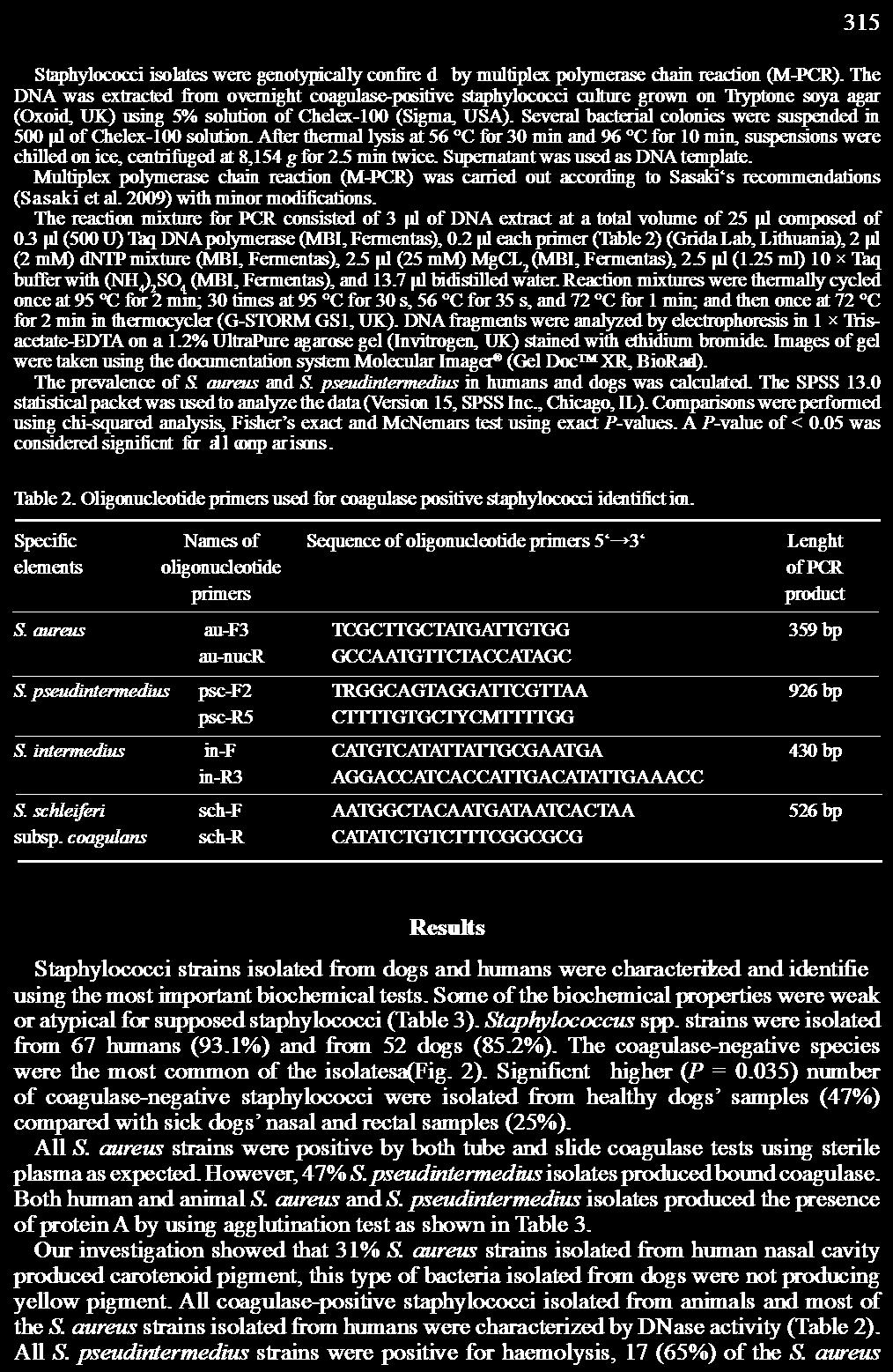
93 93
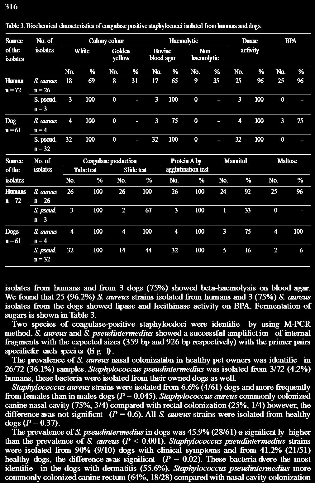
94 94
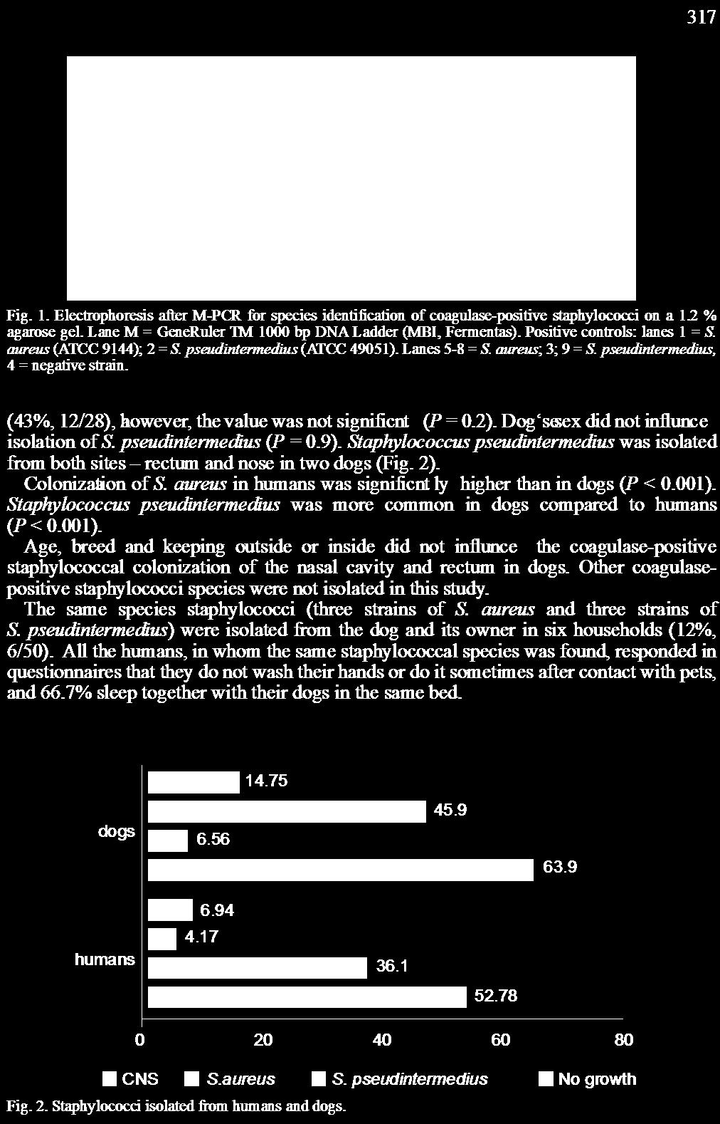
95 95
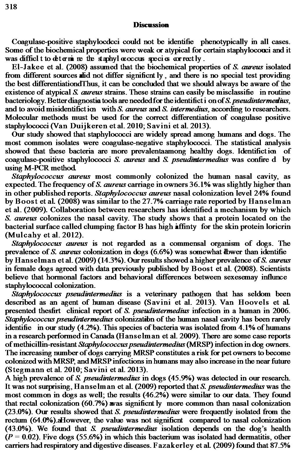
96 96
97 97
98 98
99 99
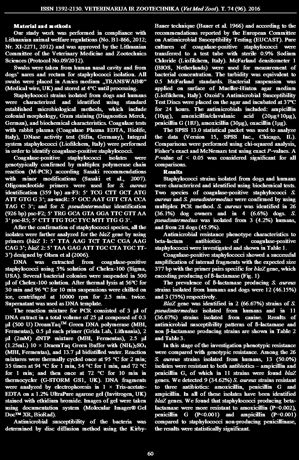
100 100
101 101

102 102
103 103
104 SANTRAUKA ĮVADAS Šunys ir katės yra vieni populiariausių gyvūnų augintinių Europoje. Pagal 2017 metais pateiktus statistikos duomenis, apie 21 proc. žmonių augina šunis, o 24 proc. kates. Bendravimo ypatumai tarp naminių gyvūnų ir žmonių per pastaruosius metus yra pasikeitę. Anksčiau šunys ir katės buvo laikomi lauke, šiandien jie dažniausiai laikomi namuose. Pastebimas glaudus fizinis kontaktas tarp gyvūno ir jo šeimininko: dažnas glostymas, glaudimas prie savęs, žmonės neretai gyvūnams leidžia laižyti savo veidą ar rankas, miegoti savo lovoje, maudytis toje pačioje vonioje ir t. t. Tačiau nereikėtų pamiršti, kad gyvūnai augintiniai ir žmonės gali būti įvairių bakterijų, tarp jų ir patogeninių bei antimikrobinėms medžiagoms atsparių bakterijų, nešiotojai. Vienos labiausiai paplitusių patogeninių bakterijų stafilokokai. Tai didžiulės klinikinės ir biotechnologinės svarbos gramteigiamos, sferinės bakterijos. Stafilokokinės infekcijos yra dažnos veterinarinėje ir žmonių medicinoje. Visame pasaulyje stafilokokinėms infekcijoms gydyti dažniausiai yra naudojami beta-laktaminiai antibiotikai. Atsparumas antimikrobinėms medžiagoms kinta ir paprastai didėja dažnai naudojamoms antimikrobinėms medžiagoms. Pastaraisiais dešimtmečiais daugelyje šalių sparčiai auga meticilinui, oksacilinui, bei kitiems beta-laktaminiams antibiotikams atsparių stafilokokų padermių skaičius. Meticilinui atsparūs patogeniniai stafilokokai aptinkami ir visuomenėje. Jeigu toks mikroorganizmas patenka į sergančio ar nusilpusio imuniteto žmogaus organizmą, gali sukelti žaizdų infekciją, sepsį, plaučių uždegimą. Literatūroje pateikiami duomenys, kad meticilinui atsparių patogeninių stafilokokų padermės, išskirtos iš šunų ir kačių, yra identiškos arba artimos žmonių epideminei hospitalinei tų šalių MASA (meticilinui atsparių Staphylococcus aureus) infekcijai. Taip pat literatūroje nurodoma, kad šie mikroorganizmai plinta ne tik tarp smulkiųjų gyvūnų, bet ir tarp juos gydančių veterinarijos gydytojų, bei augintinių savininkų. Tikslas: Nustatyti Staphylococcus aureus ir Staphylococcus pseudintermedius nešiojimo mastą gyvūnų augintinių ir jų savininkų populiacijoje bei atsparumą antimikrobinėms medžiagoms. 104
105 Uždaviniai: 1. Išskirti ir identifikuoti genotipiniais tyrimo metodais Staphylococcus aureus bei Staphylococcus pseudintermedius bakterijas gyvūnų augintinių ir jų savininkų populiacijoje. 2. Įvertinti rizikos veiksnius, kuriems esant gyvūnai augintiniai ir jų savininkai gali tapti Staphylococcus aureus ir Staphylococcus pseudintermedius nešiotojais. 3. Išskirtoms Staphylococcus aureus ir Staphylococcus pseudintermedius padermėms nustatyti filogenetinius ryšius. 4. Įvertinti išskirtų koaguliazei teigiamų stafilokokų atsparumo antimikrobinėms medžiagoms ypatumus. Praktinė reikšmė ir mokslinis naujumas Staphylococcus pseudintermedius buvo išskirta kaip nauja patogeninė stafilokokų rūšis palyginti neseniai mūsų šalyje nėra pakankamai duomenų apie šių bakterijų atsparumą meticilinui ir kitoms antimikrobinėms medžiagoms. Kadangi veterinarijos klinikose dažnai taikomas empirinis gydymas, antimikrobiniai preparatai yra naudojami siekiant išvengti antrinės infekcijos naudinga žinoti, kurie antimikrobiniai preparatai geriausiai veikia patogeninius stafilokokus. Šiais laikais yra populiaru auginti vieną ar net keletą naminių gyvūnėlių. Glaudus ryšys tarp augintinių ir jų savininkų yra dažnas. Būtina ištirti veiksnius, kurie gali turėti įtakos patogeninių bakterijų perdavimui tarp augintinių ir jų šeimininkų. Tyrimo metu nustatyta Staphylococcus pseudintermedius padermių kolonizacija ne tik gyvūnams augintiniams, bet ir žmonėms. Tai yra vertintina kaip zoonozė. Nustatytos Staphylococcus aureus ir Staphylococcus pseudintermedius padermės, turinčios blaz geną, koduojantį atsparumą beta-laktaminiams antibiotikams. Toms pačioms Staphylococcus aureus ir Staphylococcus pseudintermedius padermėms, išskirtoms iš žmonių ir šunų, nustatyti filogenetiniai ryšiai. MEDŽIAGOS IR METODAI Tyrimai atlikti Lietuvos sveikatos mokslų universiteto Veterinarijos akademijos Veterinarinės Patobiologijos katedros (buvusi Užkrečiamųjų ligų katedra) Mikrobiologijos laboratorijoje bei VDU Gamtos mokslų fakulteto Biologijos katedroje. Tyrimai atlikti laikantis Lietuvos gyvūnų gerovės taisyklių (Nr. B1-866, 2012; Nr. XI-2271, 2012) bei patvirtinti Lietuvos veterinarinės medicinos ir zootechnikos mokslų komiteto (protokolas Nr. 09/2012). 105
106 Mėginių ėmimas laboratoriniams tyrimams Prieš atliekant tyrimą, dalyvaujantiems tyrime asmenims buvo pateikta asmens, dalyvaujančio mikrobiologiniame tyrime, informavimo forma. Informuotas asmuo savo ir gyvūno augintinio dalyvavimą tyrime patvirtino pasirašydamas sutikimą. Asmenys, sutikę dalyvauti tyrime, užpildė anketą apie savo augintinį (rūšis, veislė, amžius, lytis), bendravimo įpročius su gyvūnu bei kitus duomenis, galinčius turėti įtakos patogeninių stafilokokų perdavimui tarp žmonių ir gyvūnų augintinių (laikymo sąlygos, kontaktas su kitais gyvūnais ir pan.). Gyvūnų augintojai, dalyvavę tyrime, tiriamąją medžiagą iš savo nosies steriliu vatos tamponėliu ėmė patys. Tamponas buvo įvedamas į kiekvieną nosies landą 1 2 cm gylyje, sukamas palei nosies ertmės gleivinę, išimamas ir dedamas į transportinę terpę. Gyvūnams augintiniams buvo imami du mėginiai: iš tiesiosios žarnos, įvedus sterilų tamponėlį apie 1cm į tiesiąją žarną, ir iš nosies, įvedus sterilų tamponėlį apie 0,5 1 cm į kiekvieną šnervę. Visi mėginiai iš žmonių nosies ertmės ir augintinių nosies ertmės bei tiesiosios žarnos buvo imami steriliais tamponais ir talpinami į sterilias transportines terpes TRANSWAB (Amies, Liofilchem, Italija). Iki pristatymo į laboratoriją mėginiai buvo laikomi +4 C temperatūroje. Pristatomi per dvi dienas kartu su užpildytomis anketomis ir sutikimais. Stafilokokų išskyrimas Tiriamoji medžiaga buvo sėjama į triptono sojos sultinį (angl. Tryptone Soya Broth, Oxoid, Anglija), kultivuota aerobinėmis sąlygomis 24 val. +37 C temperatūroje. Po kultivavimo bakterijų kultūra persėta ant selektyvinės ir diferencinės-diagnostinės mitybinės terpės manito druskos agaro (angl. Mannitol Salt Phenol Red Agar, Sigma, USA) stafilokokams išskirti ir manito skaidymui nustatyti. Gryna stafilokokų kultūra, persėta ant triptono sojos agaro, po kultivavimo vertintos kultūrinės savybės pigmento stafiloksantino gamyba. Mikroorganizmų morfologijai nustatyti paruošti tepinėliai iš išaugusių mikroorganizmų kultūrų, dažyti Gramo metodu (Gram, 1884) ( Diagnostica Merck, Vokietija) ir vertinti mikroskopu. Stafilokokai nuo streptokokų atskirti katalazės testu naudojant vandenilio peroksidą. Staphylococcus spp. genties nuo Micrococcus spp. genties atskyrimui naudotas greitasis testas Oksidase test stick (Liofilchem, Italija). Hemolizinėms savybėms nustatyti, stafilokokų kultūra sėta ant kraujo agaro (5 proc. galvijo kraujo ir 106
107 triptono sojos agaras) ir kultivuota val. +37 C temperatūroje. Surištoji koaguliazė nustatyta paviršiaus koaguliazės testu, laisvoji koaguliazė koaguliazės testu mėgintuvėlyje naudojant Coagulase Plasma EDTA liofilizuotą triušio plazmą (Biolife, Italija). Nustatyti, ar tiriamasis mikroorganizmas sintetina fermentą deoksiribonukleazę, atliktas DNazės testas, naudojant DNAase agarą (DNase-Agar, Sifin, Germany) ir 1N druskos rūgštį. Lipazei ir lecitinazei nustatyti stafilokokų kultūra sėta ant Baird- Parker agaro (angl. Baird-Parker medium, Liofilchem, Italija). Maltozės skaidymui nustatyti naudotas Purple agar base (Liofilchem, Italija) agaras su 1 proc. maltoze. Acetoino gamyba nustatyta Voges-Proskauer testu (Barritt, 1936). Išskirtų stafilokokų proteinui A ir rišamajam (klampumo) faktoriui nustatyti, atliktas Staphytest Plus testas (Oxoid, Anglija). Staphylococcus aureus ir Staphylococcus pseudintermedius genotipinis identifikavimas Stafilokokų DNR išskirti naudotas 5 proc. Chelex-100 tirpalas (Sigma, JAV). Išskirti koaguliazei teigiami stafilokokai iki rūšies identifikuoti naudojant daugkartinę polimerazės grandininę reakciją (angl. multiplex-pcr). Šia reakcija vienu metu galima naudoti keletą skirtingų oligonukleotidinių pradmenų ir amplifikuoti keletą skirtingų DNR sekų. Su išskirta stafilokokų padermių DNR buvo ruošiamas 25 μl PGR mišinys: 3 μl tiriamosios DNR, 0,3 μl (500 TV) Taq DNR polimerazės (MBI, Fermentas), po 0,2 μl kiekvieno oligonukleotidinių pradmenų (Grida Lab, Lietuva), 2 μl (2 mm) dntp mišinio (MBI, Fermentas), 2,5 μl (25 mm) MgCL 2 (MBI, Fermentas), 2,5 μl (1,25 ml) 10 Taq buferio su (NH 4 ) 2 SO 4 (MBI, Fermentas) ir 13,7 μl bidistiliuoto vandens. PGR 32 ciklų amplifikacija atlikta termocikleryje (G-STORM GS1, UK). Dažyti 1,3 proc. etidžio bromidu amplifikuoti mėginiai buvo analizuojami elektroforeze 1 Tris-acetate-EDTA 1,2 proc. agarozės gelyje (UltraPure agarose, Invitrogen). DNR fragmentų vaizdas gelyje buvo gautas ir nufotografuotas naudojant dokumentacijos sistemą Molecular Imager Gel Doc XR, BioRad. Stafilokokai buvo identifikuoti atsižvelgiant į kiekvienai stafilokokų rūšiai būdingą oligonukleotidinių pradmenų bazių porų skaičių genome: S. aureus 359 bp, S. pseudintermedius 926 bp, S. intermedius 430 bp, S. schleiferi subsp. coagulans 526 bp. Atsparumo antimikrobinėms medžiagoms nustatymas S. aureus ir S. pseudintermedius atsparumas antimikrobinėms medžiagoms nustatytas pagal modifikuotą Kirby-Bauer metodą (Bauer ir kiti, 107
108 1966), laikantis The European Committee on Antimicrobial Susceptibility Testing (EUCAST, 2017) standartų. Tiriamųjų mikroorganizmų ir 0.9 proc. druskos rūgšties suspensija, atitinkanti 0,5 McFarland standartą, sėta ant specialios Mueller Hinton Agar terpės (Oxoid, Anglija), ant agaro paviršiaus dėti antimikrobinių medžiagų diskai. Studijos metu atsparumas tirtas šioms antimikrobinėms medžiagoms: ampicilinui (10 μg), amoksicilinui su klavulano rūgštimi (20 μg + 10 μg), penicilinui G (1 μtv), amoksicilinui (30 μg), oksacilinui (1 μg), tilozinui, (30 μg), neomicinui (30 μg), fuzidino rūgščiai (10 μg), gentamicinui (10 μg), klindamicinui (2 μg), klaritromicinui (15 μg), enrofloksacinui (15 μg), linkomicinui (2 μg), doksiciklinui (10 μg), cefaleksinui (30 μg), cefovecinui (30 μg), cefaklorui (30 μg). Atsparumas antimikrobinėms medžiagoms vertintas po 18±2 val. kultivavimo +35±1 C temperatūroje bakterijų augimo slopinimo zoną: jautru arba atsparu. Etaloninės S. aureus (ATCC 9144) ir S. pseudintermedius (ATCC 49051) padermės buvo naudojamos antimikrobinio atsparumo tyrimo kokybės kontrolei. Atsparumo genų nustatymas Polimerazės grandinine reakcija (PGR) koagulazei teigiamų stafilokokų padermėms buvo tirti atsparumo genai, koduojantys atsparumą beta-laktamams (blaz) ir meticilinui (meca). Iš tiriamųjų stafilokokų padermių DNR buvo ruošiamas 25 μl PGR mišinys: 3 μl tiriamosios DNR, 0,3 μl (500 TV) DreamTaq TM Green DNA polymerase (MBI, Fermentas), po 0,2 μl oligonukleotidinių pradmenų (Grida Lab, Lietuva), 2 μl (2 mm) dntp mišinio (MBI, Fermentas), 2,5 μl (25 mm) MgCL 2 (MBI, Fermentas), 2.5 μl (1.25mL) 10 DraemTaq Green buferio su (NH 4 ) 2 SO 4 (MBI, Fermentas) ir 13,7 μl bidistiliuoto vandens. PGR 37 ciklų amplifikacija atlikta termocikleryje (G-STORM GS1, UK). Amplifikuotieji mėginiai buvo analizuojami elektroforeze 1 Tris-acetate- EDTA 1,2 proc. agarozės gelyje (UltraPure agarose, Invitrogen). DNR fragmentų vaizdas gelyje buvo gautas ir nufotografuotas naudojant dokumentacijos sistemą Molecular Imager Gel Doc XR, BioRad. Atsparumo genai buvo nustatomi atsižvelgiant į kiekvienam genui būdingą oligonukleotidinių pradmenų bazių porų skaičių genome: beta-laktamams (blaz) 377 bp ir meticilinui (meca). 108
109 Staphylococcus aureus ir Staphylococcus pseudintermedius nuc geno sekvenavimas Tais atvejais, kai buvo nustatyta ta pati stafilokokų rūšis žmogui ir jo augintiniui, buvo atliktas nuc geno sekvenavimas Sangerio metodu. Filogenetiniams ryšiams nustatyti naudota kompiuterinė programa Mega 7 (Tamura ir kt., 2016). Sekoskaitos nukleotidų analizė atlikta Neighbor-Join metodu (Saitou, Nei, 1987). Filogenetinis medis su nukleotidais sudarytas remiantis Kimura 2-parameter modeliu (Kimura, 1980). Gautos genų sekos buvo palygintos tarpusavyje ir su atitinkamomis stafilokokų sekomis, gautomis iš tarptautinio genų banko duomenų. Statistinė analizė Statistinė analizė atlikta naudojant SPSS 13.0 statistinį paketą (15 versija, SPSS Inc., Chicago, IL). Išskirtų mikroorganizmų veiksniai ir aplinkybės, galinčios turėti įtakos jų buvimui (šaltinis, nešiotojo amžius, lytis kt.), buvo lyginami naudojant Chi-kvadrato analizę, tikslų Fisher'ą ir McNemars testą naudojant tikslias P reikšmes. Visų palyginimų atveju reikšmės p< 0,05 laikytos reikšmingomis. 95 proc. pasikliautinas intervalas (PI 95 proc.) buvo apskaičiuotas naudojant StatPages.net (Pezzullo, 2009) kompiuterinę programą. Tiriamųjų apžvalga REZULTATAI Bakterijų mėginiai buvo paimti iš 61 šuns, 45 kačių ir 107 gyvūnų augintojų. Iš viso buvo ištirti 122 šunų ir 90 kačių nosies ertmės ir tiesiosios žarnos mėginiai, bei 107 žmonių nosies ertmės mėginiai, siekiant išskirti koaguliazei teigiamus stafilokokus. Mėginiai bakteriologiniam tyrimui buvo paimti iš skirtingo amžiaus ir lyties, įvairių veislių, sveikų bei sergančių šunų ir kačių. Sergančių gyvūnų dažniausi klinikiniai sutrikimai buvo: dermatitas, kvėpavimo takų ir virškinimo trakto ligos. Gydymas antimikrobiniais preparatais, prieš imant mėginius, buvo taikytas devyniems šunims ir keturioms katėms. Dažniausiai gydymui buvo naudotos antimikrobinės medžiagos: amoksiciklinas su klavulano rūgštimi, gentamicinas arba enrofloksacinas. Iš visų tyrime dalyvavusių naminių gyvūnėlių savininkų 72 augino bent vieną šunį, 48 žmonių namuose buvo auginama bent viena katė. 109
110 Visi 107 tyrimo dalyviai buvo Lietuvos gyventojai; vidutinis dalyvių amžius buvo 34 metai (apimtis metų). Tyrime dalyvavo 77,6 proc. moterų ir 22,4 proc. vyrų. Septyni naminių gyvūnėlių savininkai (6,5 proc.) dirbo sveikatos priežiūros srityje ligoninėje. Aštuoniolika (16,8 proc.) buvo veterinarijos studentai. 57 (79,2 proc.) šunų šeimininkai atsakė, kad tarp jų ir šuns yra glaudus ryšys jie savo gyvūną glaudžia prie savęs ir glosto, o 15 respondentų atsakė beveik neturintys kontakto su savo augintiniu. 35 (48,6 proc.) tyrimo dalyviai atsakė, kad leidžia savo šuniui ilsėtis arba miegoti savo lovoje; visi atsakė, kad leidžia šuniui laižyti jų rankas, o 34 (47,2 proc.) veidą. 29 (40,3 proc.) šunų savininkai anketoje nurodė, kad po sąlyčio su šunimi jie visada nusiplauna rankas. 7 (9,7 proc.) dalyviai teigė, kad valgo su savo šunimis iš tų pačių indų. Iš 43 kačių savininkų, dalyvavusių apklausoje, 97,7 proc. teigė, kad jie kontaktuoja su savo gyvūnais dažnai glosto ir glaudžia prie savęs. 37 (86,0 proc.) dalyviai nurodė, kad leidžia savo katėms miegoti savo lovoje, 25 proc. (58,1 proc.) leidžia katėms laižyti rankas, 10 proc. (23,3 proc.) laižyti veidą. 9 (20,9 proc.) kačių savininkai nurodė neplaunantys rankų po sąlyčio su kate. Staphylococcus aureus ir Staphylococcus pseudintermedius išskyrimas ir pagrindinės savybės Visi tiriamieji stafilokokai buvo gramteigiami kokai, sintetino katalazę, bei buvo neigiami oksidazės testui. Po 24 val. kultivavimo +37 C temperatūroje triptono sojos agare, 31 proc. Staphylococcus aureus padermių, išskirtų iš žmogaus nosies ertmės, gamino geltonos spalvos karotinoidinį pigmentą stafiloksantiną. Visos Staphylococcus aureus ir Staphylococcus pseudintermedius padermės koaguliavo plazmą mėgintuvėlyje po inkubavimo +37 C temperatūroje. Atlikus paviršiaus koaguliazės testą, naudojant sterilią triušio plazmą, agliutinacija pasireiškė visose Staphylococcus aureus padermėse, o surištoji koaguliazė buvo nustatyta ir daugelyje Staphylococcus pseudintermedius padermių, išskirtų iš šunų (44,0 proc.), iš žmonių (67,0 proc.) ir iš kačių (100 proc.). Visos Staphylococcus aureus ir Staphylococcus pseudintermedius padermės agliutinavo, naudojant greitąjį baltymo A ir rišamojo (klampumo) faktoriaus testą. Dalis Staphylococcus aureus padermių, nustatytų žmonėms (65 proc.) ir šunims (75 proc.), sukėlė beta-hemolizę kraujo agare po inkubacijos 24 val. +37 C temperatūroje. Visos Staphylococcus pseudintermedius padermės 110
111 suformavo dvigubą hemolizės zoną kraujo agare po 24 val. inkubacijos +37 C temperatūroje. 25 (96,2 proc.) Staphylococcus aureus padermės, išskirtos iš žmonių, 3 (75 proc.) iš šunų ir 3 (100 proc.) iš kačių, parodė lipazės ir lecitinazės aktyvumą ant BPA. Pridėjus 1 N vandenilio chlorido rūgšties ant DNase agaro su kultivuotomis stafilokokų kultūromis, dezoksiribonukleazės aktyvumas skaidri zona aplink stafilokokų kultūrą buvo matoma visose koaguliazei teigiamų stafilokokų padermėse, išskirtose iš gyvūnų augintinių, ir daugumoje Staphylococcus aureus padermių, izoliuotų nuo žmonių. Didžioji dalis Staphylococcus aureus bakterijų, išskirtų iš žmonių (94 proc.), šunų (75 proc.) ir kačių (100 proc.), skaidė manitolį. Keletas Staphylococcus pseudintermedius padermių fermentavo manitolį taip pat: 1 (33 proc.) iš žmonių ir 5 (16 proc.) iš šunų. Maltozę skaidė 30 (91 proc.) Staphylococcus aureus padermių, išskirtų iš žmonių, ir visos iš gyvūnų augintinių. Dvi (6 proc.) Staphylococcus pseudintermedius padermės iš šunų fermentavo maltozę taip pat. Išanalizavus išskirtų koaguliazei teigiamų stafilokokų kultūrines ir biochemines savybes, virulentiškumo veiksnius, 85 proc. Staphylococcus aureus ir 52,9 proc. Staphylococcus pseudintermedius padermių šios savybės buvo tipinės. Kadangi kai kurios biocheminės savybės buvo sunkiai vertinamos arba netipinės tiriamiems stafilokokams, o Staphylococcus intermedius grupės (S. intermedius, S. delphini ir S. pseudintermedius) rūšies bakterijų negalima patikimai atskirti viena nuo kitos naudojant fenotipinius tyrimus arba komercinius diagnostinius rinkinius, buvo atlikti genetiniai tyrimai. Daugkartinė polimerazės grandininė reakcija buvo naudojama siekiant tiksliai identifikuoti koaguliazei teigiamus stafilokokus. Gauti specifiniai DNR fragmentų amplifikavimo 359 bp ir 926 bp produktai, kurių dydis atitiko dviejų stafilokokų rūšių: S. aureus ir S. pseudintermedius genomą. Staphylococcus aureus ir Staphylococcus pseudintermedius paplitimas Staphylococcus spp. bakterijos buvo išskirtos iš 102 (95,3 proc.) žmonių, 52 (85,2 proc.) šunų ir iš 43 (95,6 proc.) kačių. Nustatytos koaguliazei teigiamų stafilokokų rūšys pateiktos 1 paveikslėlyje. 111
. Šios rūšies stafilokokų kolonizacija buvo didesnė tarp žmonių nei tarp šunų ar kačių (p < 0,001). Staphylococcus pseudintermedius buvo išskirtas iš 3 žmonių (2,8 proc.")
112 1 pav. Stafilokokų paplitimas tarp žmonių, šunų ir kačių (proc.) Staphylococcus aureus buvo nustatytas 33 žmonėms (30,8 proc.; PI 95 proc. 22,9 40,1), 4 šunims (6,6 proc.; PI 95 proc. 2,6 15,7) ir 3 katėms (6,7 proc.; PI 95 proc. 2,3 17,9). Šios rūšies stafilokokų kolonizacija buvo didesnė tarp žmonių nei tarp šunų ar kačių (p < 0,001). Staphylococcus pseudintermedius buvo išskirtas iš 3 žmonių (2,8 proc.; PI 95 proc. 1 7,9), 28 šunų (45,9 proc.; PI 95 proc. 34,0 58,3) ir 1 katės (2,2 proc.; PI 95 proc. 0,4 11,6). Staphylococcus pseudintermedius padermės buvo išskirtos dažniau iš šunų, sergančių odos ligomis, palyginti su sveikais šunimis (p = 0,03). Ši stafilokokų rūšis šunims buvo nustatyta dažniau nei žmonėms ir katėms (p < 0,001). Staphylococcus aureus ir Staphylococcus pseudintermedius atsparumas antimikrobinėms medžiagoms Studijos metu Staphylococcus aureus ir Staphylococcus pseudintermedius padermėms buvo nustatytas atsparumas: penicilinams, makrolidams, linkozamidams, chinolonams, trijų kartų cefalosporinams ir tetraciklinams. 45,4 proc. Staphylococcus pseudintermedius padermių buvo atsparios penicilinui G ir ampicilinui, 29,0 proc. amoksicilinui, 26,7 proc. klindamicinui ir klaritromicinui, 23,3 proc. doksiciklinui ir tilozinui. 29,0 proc. S. pseudintermedius buvo jautrūs visoms antimikrobinėms medžiagoms. Septyni šunys (11,5 proc.; PI 95 proc. 5,7 21,8) buvo Staphylococcus pseudintermedius pasižyminčių dauginiu atsparumu, nešiotojai (atsparumas 3 ar daugiau klasių antimikrobinėms medžiagoms). 67,5 proc. Staphylococcus aureus padermių buvo atsparios ampicilinui, 62,5 proc. penicilinui G, 42,5 proc. amoksicilinui, 15,0 proc. tilozinui. 32,5 proc. Staphylococcus aureus padermių buvo jautrios visoms antimikrobinėms medžiagoms. Vienai Staphylococcus aureus padermei (1,6 proc.; 112
AB Linas Agro Group 2018 m. spalio 31 d. eilinio visuotinio akcininkų susirinkimo BENDRASIS BALSAVIMO BIULETENIS GENERAL VOTING BALLOT at Annual Gener
 1 AKCININKAS SHAREHOLDER FIZINIS ASMUO / NATURAL PERSON Vardas, pavardė Name, surname Asmens kodas: Personal code: JURIDINIS ASMUO / LEGAL PERSON Juridinio asmens pavadinimas Name of legal person Juridinio
Priedai
 Priedai Priedas Nr. 3 Įvesti duomenys Na- smūgių dažnumas į 1km' Na= 2 v 4 4 C2= 1 - objekto konstrukcija L- objekto ilgis L= 24 C3= 1 - objekto vertė W- objekto plotis W= 12 C4= 1 - žmonių kiekis objekte
Civilinės aviacijos administracija
 SAFA programa. Aktualūs klausimai Andrejus Golubevas Skrydžių priežiūros skyriaus vyriausiasis specialistas 2012-11-12 1 Prezentacijos turinys SAFA programos teisinis pagrindas SAFA programos principai
NEPRIKLAUSOMO AUDITORIAUS IŠVADA UAB Vilniaus gatvių apšvietimo elektros tinklai akcininkui Nuomonė Mes atlikome UAB Vilniaus gatvių apšvietimo elektr
 NEPRIKLAUSOMO AUDITORIAUS IŠVADA UAB Vilniaus gatvių apšvietimo elektros tinklai akcininkui Nuomonė Mes atlikome UAB Vilniaus gatvių apšvietimo elektros tinklai (toliau Įmonė) finansinių ataskaitų, kurias
AB Linas Agro Group 2018 m. spalio 31 d. eilinio visuotinio akcininkų susirinkimo BENDRASIS BALSAVIMO BIULETENIS GENERAL VOTING BALLOT at Annual Gener
 AKCININKAS THE SHAREHOLDER FIZINIS ASMUO / NATURAL PERSON Vardas, pavardė Name, surname Asmens kodas Personal code JURIDINIS ASMUO / LEGAL PERSON Juridinio asmens pavadinimas Name of legal person Juridinio
Vienlusčių įtaisų projektavimas
 Vienlusčių įtaisų projektavimas 5 paskaita Baigtiniai būsenų automatai Baigtinis būsenų automatas: angl. Finite State Machine (FSM) FSM yra nuosekliai būsenas keičiantis automatas su "atsitiktine" kitos
CDK4/6 kelio aktyvacija onkologijoje, molekulinių biomarkerių detekcija ir jų inhibicijos biologija Vaidas Dirsė, Rimvydas Norvilas
 CDK4/6 kelio aktyvacija onkologijoje, molekulinių biomarkerių detekcija ir jų inhibicijos biologija Vaidas Dirsė, Rimvydas Norvilas CDK nuo ciklinų priklausoma kinazė Ciklinai CKI Ciklinų kinazių inhibitoriai
Programų sistemų inžinerija Saulius Ragaišis, VU MIF
 Programų sistemų inžinerija 2014-02-12 Saulius Ragaišis, VU MIF saulius.ragaisis@mif.vu.lt SWEBOK evoliucija Nuo SWEBOK Guide to the Software Engineering Body of Knowledge, 2004 Version. IEEE, 2004. prie
Microsoft Word - AIKOS IKUP1
 STUDIJŲ PROGRAMA. IKIMOKYKLINIO UGDYMO PEDAGOGIKA Parametrai Studijų programos pavadinimas Ikimokyklinio ugdymo pedagogika Studijų programos valstybinis kodas 653X11005 Studijų programos kodas pagal Tarptautinę
rp_ IS_2
 Page 1 of 5 Pagrindinė informacija Juodoji aromatizuota arbata su miško uogomis Exemption Flags Exempt from Artwork Exempt from NEP Reporting Produkto pavadinimas Country Lipton Brand Name Forest Fruits
DARBO SUTARTIS
 PATVIRTINTA Lietuvos Respublikos švietimo ir mokslo ministro 2016 m. lapkričio 16 d. įsakymu Nr. V-1011 (Studento praktinio mokymo sutarties pavyzdinė forma) STUDENTO PRAKTINIO MOKYMO PAVYZDINĖ SUTARTIS
Vilius Grabauskas Jūratė Klumbienė Janina Petkevičienė Edita Šakytė Vilma Kriaučionienė Aurelijus Veryga Ritva Prättälä SUAUGUSIŲ LIETUVOS ŽMONIŲ GYVE
 Vilius Grabauskas Jūratė Klumbienė Janina Petkevičienė Edita Šakytė Vilma Kriaučionienė Aurelijus Veryga Ritva Prättälä SUAUGUSIŲ LIETUVOS ŽMONIŲ GYVENSENOS TYRIMAS, 2008 Health Behaviour among Lithuanian
Ekonomikos inžinerija, Globalioji ekonomika NR. Baigiamojo darbo temos pavadinimas Baigiamojo darbo vadovas, kontaktai 1. Globalizacijos poveikis X se
 Ekonomikos inžinerija, Globalioji ekonomika NR. Baigiamojo darbo temos pavadinimas Baigiamojo darbo vadovas, kontaktai 1. Globalizacijos poveikis X sektoriaus įmonių specializacijos lygiui. Impact of Globalization
AVK SUPA PLUS COUPLING 621/61 Tensile, for PE and PVC pipes, NF approved EPDM sealing 001 AVK Supa Plus is a range of tensile couplings, flange adapto
 AVK SUPA PUS COUPING 1/61 Tensile, for PE and PVC pipes, NF approved EPM sealing AVK Supa Plus is a range of tensile couplings, flange adaptors and end caps dedicated for PE and upvc pipes. Supa Plus couplings
Microsoft Word - Gramneigiamų-monobakteriemijų-sukėlėjų-jautrumas-antibiotikams-sutrumpinta (2).docx
.docx") Medical sciences (2018) 1 8 Gramnegative rod monobacteremia pathogens and their sensitivity to antibiotics in intensive care unit Dalia Adukauskienė 1, Juozas Žilinskas 2, Dovilė Valančienė 1 1 Lithuanian
Medical sciences (2018) 1 8 Gramnegative rod monobacteremia pathogens and their sensitivity to antibiotics in intensive care unit Dalia Adukauskienė 1, Juozas Žilinskas 2, Dovilė Valančienė 1 1 Lithuanian
SPECIALIOSIOS UŽDAROJO TIPO PRIVATAUS KAPITALO INVESTICINĖS BENDROVĖS INVL TECHNOLOGY AUDITO KOMITETO NUOSTATAI BENDROJI DALIS 1. Šie specialiosios už
 SPECIALIOSIOS UŽDAROJO TIPO PRIVATAUS KAPITALO INVESTICINĖS BENDROVĖS INVL TECHNOLOGY AUDITO KOMITETO NUOSTATAI BENDROJI DALIS 1. Šie specialiosios uždarojo tipo privataus kapitalo investicinės bendrovės
Vilius Grabauskas Jūratė Klumbienė Janina Petkevičienė Edita Šakytė Vilma Kriaučionienė Aurelijus Veryga SUAUGUSIŲ LIETUVOS ŽMONIŲ GYVENSENOS TYRIMAS,
 Vilius Grabauskas Jūratė Klumbienė Janina Petkevičienė Edita Šakytė Vilma Kriaučionienė Aurelijus Veryga SUAUGUSIŲ LIETUVOS ŽMONIŲ GYVENSENOS TYRIMAS, 2012 Health Behaviour among Lithuanian Adult Population,
Microsoft Word - Deposits and withdrawals policy 400.doc
 DEPOSITS AND WITHDRAWALS POLICY / LĖŠŲ ĮDĖJIMO IR NUĖMIMO POLITIKA TeleTrade-DJ International Consulting Ltd 2011-2017 TeleTrade-DJ International Consulting Ltd. 1 Bank Wire Transfers: When depositing
Register your product and get support at Indoor wireless headphones SHC8535 SHC8575 LT Vartotojo vadovas
 Register your product and get support at www.philips.com/welcome Indoor wireless headphones SHC8535 SHC8575 LT Vartotojo vadovas SHC8535 SHC8535 A a b B a c d b e f c C D E F a G b H I 1 Kas yra rinkinyje
A-M-K_nr-10_maketas_I.indd
 12 ISSN 1822-1645 http://dx.doi.org/10.7720/1822-1645.10.1 Dortmundo technikos universitetas, Twente universitetas Technical University of Dortmund, University of Twente Studentai kaip kokybės užtikrinimo
LIETUVOS AGRARINĖS EKONOMIKOS INSTITUTAS
 LITHUANIAN INST ITUTE OF AGRARI AN ECONOMICS LITHUANIAN FAMILY FARM PROFITABILITY: THE ECONOMIC DIMENSION OF SUSTAINABILITY SCIENTIFIC STUDY 218 Tomas BALEŽENTIS, Virginia NAMIOTKO, Lina NOVICKYTĖ Baležentis,
VILNIUS GEDIMINAS TECHNICAL UNIVERSITY Vytautas DUMBLIAUSKAS DEVELOPMENT AND APPLICATION OF TOUR-BASED TRAVEL DEMAND MODEL FOR PLANNING OF URBAN TRANS
 VILNIUS GEDIMINAS TECHNICAL UNIVERSITY Vytautas DUMBLIAUSKAS DEVELOPMENT AND APPLICATION OF TOUR-BASED TRAVEL DEMAND MODEL FOR PLANNING OF URBAN TRANSPORT NETWORKS DOCTORAL DISSERTATION TECHNOLOGICAL SCIENCES,
Slide 1
 Horizontas 2020 teminės srities Mokslas su visuomene ir visuomenei 2018 2020 m. darbo programa ir kvietimų tematika Horizontas 2020 teminės srities Mokslas su visuomene ir visuomenei informacinė diena
MHAIA Estimation of Production rd Stage
 PROBABILISTIC ESTIMATION OF HASS AVOCADO PRODUCTION ESTIMATION IN THE AREA OF THE STATE OF MICHOACAN OF HASS AVOCADO PRODUCTION IN THE AREA OF THE STATE OF MICHOACAN CERTIFIED TO EXPORT TO THE USA SEASON
PowerPoint Presentation
 Programų sistemų inžinerija 2018-02-07 Saulius Ragaišis, VU MIF saulius.ragaisis@mif.vu.lt Klausytojai: Susipažinimas Išklausyti programų sistemų inžinerijos kursai Profesinė patirtis Dabar klausomi pasirenkami
ALEKSANDRO STULGINSKIO UNIVERSITETAS LIETUVOS AGRARINIŲ IR MIŠKŲ MOKSLŲ CENTRAS Viktorija Vaštakaitė METABOLITŲ IR MINERALINIŲ ELEMENTŲ VALDYMO BRASSI
 ALEKSANDRO STULGINSKIO UNIVERSITETAS LIETUVOS AGRARINIŲ IR MIŠKŲ MOKSLŲ CENTRAS Viktorija Vaštakaitė METABOLITŲ IR MINERALINIŲ ELEMENTŲ VALDYMO BRASSICACEAE DAIGINIUOSE FOTOFIZIOLOGINIAI ASPEKTAI DAKTARO
Viesasis Nr. 17_tirazui.p65
 MYKOLO ROMERIO UNIVERSITETAS KAUNO TECHNOLOGIJOS UNIVERSITETAS ISSN 1648 2603 VIEÐOJI POLITIKA IR ADMINISTRAVIMAS Nr. 17 Vilnius * Mykolo Romerio universiteto Leidybos centras * 2006 1 Vyriausieji redaktoriai:
A-M-K_nr-11_maketas_trump.indd
 66 ISSN 1822-1645 (Print), ISSN 2345-0258 (Online) http://dx.doi.org/10.7220/2345-0258.11.3 Fernando Cardoso Sousa, Ileana Pardal Monteiro, René Pellissier Algarvės universitetas, Algarvės universitetas,
Programos Leader ir žemdirbių mokymo metodikos centras Centre for Leader Programme and Agricultural Training Methodology LEADER ĮGYVENDINIMO APŽVALGA
 Programos Leader ir žemdirbių mokymo metodikos centras Centre for Leader Programme and Agricultural Training Methodology LEADER ĮGYVENDINIMO APŽVALGA Patirtis, pamokos, perspektyvos LEADER IMPLEMENTATION
LCON F4F PROJECT MAYO DECORATIVE 3 L IS_3 LT
 Page 1 of 10 Reason For Issue Key Words Key Word Customers / MSU Customers / MSU Customers / MSU Unilever Czech Republic/Slovakia Unilever Poland & Baltics Unilever SCE Value Manufacturers Classification
PowerPoint Presentation
 Skaitmeninės kabelinės televizijos galimybės Mindaugas Žilinskas Ryšių reguliavimo tarnybos Radijo ryšio departamento direktorius Vilnius, 2012 m. 1 Turinys 1. Raiškiosios (HD) televizijos standartai ir
Finansų inžinerijos studijų programa (6211LX060) Specializacija Baigiamojo darbo temos pavadinimas Baigiamojo darbo temos išskirtinumas ir vadovo pasi
 Specializacija Baigiamojo darbo temos pavadinimas Baigiamojo darbo temos išskirtinumas ir vadovo pasi") Finansų inžinerijos studijų programa (6211LX060) Specializacija Baigiamojo darbo temos pavadinimas Baigiamojo darbo temos išskirtinumas ir vadovo pasiekimai, siejant su siūloma tema Baigiamojo darbo vadovas
Finansų inžinerijos studijų programa (6211LX060) Specializacija Baigiamojo darbo temos pavadinimas Baigiamojo darbo temos išskirtinumas ir vadovo pasiekimai, siejant su siūloma tema Baigiamojo darbo vadovas
PR_COD_1amCom
 Europos Parlamentas 2014-2019 Piliečių laisvių, teisingumo ir vidaus reikalų komitetas 2016/0225(COD) 23.3.2017 ***I PRANEŠIMO PROJEKTAS dėl pasiūlymo dėl Europos Parlamento ir Tarybos reglamento, kuriuo
Microsoft Word asis TAS.DOC
 Tarptautinių audito ir užtikrinimo standartų valdyba 501-asis TAS 2009 balandis Tarptautinis audito standartas Audito įrodymai konkretūs svarstymai dėl atskirų sričių 1 International Auditing and Assurance
Dokumentų leidimui laikinai gyventi Lietuvos Respublikoje gauti pateikimo ir leidimų laikinai gyventi Lietuvos Respublikoje užsieniečiams išdavimo, ke
 (įstaigos pavadinimas) GAUTA Nr. PRAŠYMAS IŠDUOTI LEIDIMĄ LAIKINAI GYVENTI LIETUVOS RESPUBLIKOJE APPLICATION FOR THE ISSUE OF A TEMPORARY RESIDENCE PERMIT IN THE REPUBLIC OF LITHUANIA (data / date) Prašau
PowerPoint Presentation
 SenGor partner meeting Leuven. Lithuanian countryside tourism association activities www.countryside.lt 2015 Nov, President Linas Zabaliunas Summary of the activities Jun - Nov o Working on the draft guidelines
Slide 1
 H2020 Pažangos sklaida ir dalyvavimo plėtra Informacinis renginys, Lietuvos mokslo taryba Živilė Ruželė, zivile.ruzele@lmt.lt 2019 m. birželio 7 d. Turinys 1. Plėtros stipendijos 2. Patarimai Twinning
Neiškiliojo optimizavimo algoritmas su nauju bikriteriniu potencialiųjų simpleksų išrinkimu naudojant Lipšico konstantos įvertį
 Neiškiliojo optimizavimo algoritmas su nauju bikriteriniu potencialiųjų simpleksų išrinkimu naudojant Lipšico konstantos įvertį. Albertas Gimbutas 2018 m. birželio 19 d. Vadovas: Prof. habil. dr. Antanas
Turinys Bendrovės vadovo žodis...2 Bendrovės valdymo struktūra...4 Bendrovės pagrindinės veiklos pobūdis...6 Bendrovės 2008 metų veiklos apžvalga...8
 Turinys Bendrovės vadovo žodis...2 Bendrovės valdymo struktūra...4 Bendrovės pagrindinės veiklos pobūdis...6 Bendrovės 2008 metų veiklos apžvalga...8 Garantijų portfelio analizė...10 Rizikos valdymas...22
Visuomenės sveikata Originalūs straipsniai Meticilinui atsparaus Staphylococcus aureus valdymo būklė Lietuvos ligoninėse Rūta Bagdonaitė 1, Rolanda Va
 Meticilinui atsparaus Staphylococcus aureus valdymo būklė Lietuvos ligoninėse Rūta Bagdonaitė 1, Rolanda Valintėlienė 1, Jolanta Griškevičienė 1, Aušra Macijauskienė 2 1 Higienos institutas 2 Vilniaus
MUITINĖS DEPARTAMENTAS PRIE LIETUVOS RESPUBLIKOS FINANSŲ MINISTERIJOS BENDRO NAUDOTOJŲ VALDYMO SISTEMOS, ATITINKANČIOS EUROPOS KOMISIJOS REIKALAVIMUS,
 MUITINĖS DEPARTAMENTAS PRIE LIETUVOS RESPUBLIKOS FINANSŲ MINISTERIJOS BENDRO NAUDOTOJŲ VALDYMO SISTEMOS, ATITINKANČIOS EUROPOS KOMISIJOS REIKALAVIMUS, SUKŪRIMO VERSIJA: v0.10 Vilnius 2018 TURINYS 1 Windows
Finansinių paraiškų teikimo, jų vertinimo, lėšų skyrimo, ataskaitų teikimo ir vertinimo tvarkos aprašo
 VILNIAUS UNIVERSITETO BIOTECHNOLOGIJOS INSTITUTAS COST veiklos Nr. TD0905 Epigenetics: from Bench to Bedside ATASKAITA UŽ 2011 metus Per ataskaitinį laikotarpį (2011 metais) buvo numatyti šie susitikimai:
274 PRIEDAI K priedas. Elektroninio vartotojo gyvavimo ciklo tyrimo duomenų charakteristikos K.1 lentelė. Klausimyno dalies, skirtos elektroninio vart
 274 PRIEDAI K priedas. Elektroninio vartotojo gyvavimo ciklo tyrimo duomenų charakteristikos K.1 lentelė. Klausimyno dalies, skirtos elektroninio vartotojo gyvavimo ciklo analizei, patikimumo vertinimas
LIETUVOS RESPUBLIKOS VALSTYBINIO PATENTŲ BIURO DIREKTORIAUS
 Įsakymas netenka galios 2015-11-24: Lietuvos Respublikos valstybinis patentų biuras, Įsakymas Nr. 3R-72, 2015-11-20, paskelbta TAR 2015-11-23, i. k. 2015-18546 Dėl Lietuvos Respublikos valstybinio patentų
DB sukūrimas ir užpildymas duomenimis
 DB sukūrimas ir užpildymas duomenimis Duomenų bazės kūrimas Naujas bendrąsias DB kuria sistemos administratorius. Lokalias DB gali kurti darbo stoties vartotojasadministratorius. DB kuriama: kompiuterio
Microsoft Word - Acute Coronary Syndrome in Patients, Younger Than 60 Years. Common Characteristics of these patients.doc
 Medical sciences (208) 5 Acute coronary syndrome in patients, younger than 60 years. Common characteristics of these patients Andrius Romeika, Artūras Smirnovas Lietuvos sveikatos mokslų universitetas,
PATVIRTINTA: LASF žiedo komiteto, Originali teksto kalba - lietuvių. The original text language is Lithuanian Turinys (Content) 1. TAISYKLĖSE NAUDOJAM
 1. TAISYKLĖSE NAUDOJAM") PATVIRTINTA: LASF žiedo komiteto, Originali teksto kalba - lietuvių. Turinys (Content) 1. TAISYKLĖSE NAUDOJAMOS SĄVOKOS 3 2. BENDROSIOS NUOSTATOS 4 3. KOMANDOS, KOMANDŲ SUDARYMAS. LENKTYNIŲ DALYVIAI 5
PATVIRTINTA: LASF žiedo komiteto, Originali teksto kalba - lietuvių. Turinys (Content) 1. TAISYKLĖSE NAUDOJAMOS SĄVOKOS 3 2. BENDROSIOS NUOSTATOS 4 3. KOMANDOS, KOMANDŲ SUDARYMAS. LENKTYNIŲ DALYVIAI 5
2014 September 30. Journal of Medical Sciences, ISSN: EARLY DIAGNOSTIC FEATURES OF KIDNEY CANCER Dr. Lilija Butkevičienė 1, Rasita Zamblausk
 EARLY DIAGNOSTIC FEATURES OF KIDNEY CANCER Dr. Lilija Butkevičienė 1, Rasita Zamblauskaitė 1, Žilvinas Pavilionis 2 Lithuanian University of Health Sciences, Radiology clinics. ABSTRACT Objective: To evaluate
Monetos_1-8 taisytas indd
 LIETUVOS MONETOS LITHUANIAN COINS LIETUVOS MONETOS LITHUANIAN COINS L I E T U V O S B A N K A S 2 0 0 6 UDK 737(474.5)(091) Li301 Teksto autoriai / Text by Arûnas Dulkys, Stanislovas Sajauskas, Juozas
805-asis Tarp TAS 2009 balandis Tarptautinis audito standartas Specialūs svarstymai atskirų finansinių ataskaitų ir atskirų finansinės ataskaitos elem
 805-asis Tarp TAS 2009 balandis Tarptautinis audito standartas Specialūs svarstymai atskirų finansinių ataskaitų ir atskirų finansinės ataskaitos elementų, sąskaitų ir straipsnių auditas 1 International
2019 m. LIETUVOS RESPUBLIKOS BURIAVIMO TAURĖS REGATA BEI VLADIMIRO IR VALERIJAUS MAMONTOVŲ ATMINIMO VĖTRUNGĖS TAURĖ d. NIDA, Lietuva LEN
 2019 m. LIETUVOS RESPUBLIKOS BURIAVIMO TAURĖS REGATA BEI VLADIMIRO IR VALERIJAUS MAMONTOVŲ ATMINIMO VĖTRUNGĖS TAURĖ 2019 05 10-12 d. NIDA, Lietuva LENKTYNIŲ INSTRUKCIJA LITHUANIAN SAILING CUP REGATTA 2019
VIZOS GAVIMAS-INFO
 KAIP KREIPTIS DĖL VIZOS 1. Sumokėkite konsulinį mokestį. Kiekvieno besikreipiančio dėl vizos asmens vardu, įskaitant mažamečius vaikus ir pensininkus, turi būti sumokėtas negrąžinamas konsulinis mokestis.
Saziningumo_pasizadejimu_eskperimentas_8(a5)
") L i e t u v o s s k y r i u s KAS MOKSLEIVIUS SKATINA ELGTIS SĄŽININGAI? PRAKTINIS VADOVAS VILNIUS, 2019 Transparency International Lietuvos skyrius yra ne pelno siekianti, nepolitinė, nevyriausybinė organizacija,
L i e t u v o s s k y r i u s KAS MOKSLEIVIUS SKATINA ELGTIS SĄŽININGAI? PRAKTINIS VADOVAS VILNIUS, 2019 Transparency International Lietuvos skyrius yra ne pelno siekianti, nepolitinė, nevyriausybinė organizacija,
Dalyvavimo COST veiklos IC1201 Behavioural Types for Reliable Large-Scale Software Systems (BETTY) valdymo komiteto posėdyje ATASKAITA Vieta: Viešbuči
 valdymo komiteto posėdyje ATASKAITA Vieta: Viešbuči") Dalyvavimo COST veiklos IC1201 Behavioural Types for Reliable Large-Scale Software Systems (BETTY) valdymo komiteto posėdyje ATASKAITA Vieta: Viešbučio Grand Hotel Palatino konferencijų salė, Roma (Italija)
Dalyvavimo COST veiklos IC1201 Behavioural Types for Reliable Large-Scale Software Systems (BETTY) valdymo komiteto posėdyje ATASKAITA Vieta: Viešbučio Grand Hotel Palatino konferencijų salė, Roma (Italija)
Microsoft PowerPoint - Sveik_sist_fin_ir_pasl_priein_prezentacija_pataisyta
 Studija Lietuvos sveikatos apsaugos sistemos finansavimas ir sveikatinimo paslaugų prieinamumas gyventojams Studiją parengė UAB Sveikatos ekonomikos centras bendradarbiaujant su Etinių farmacijos kompanijų
Bioness
 Inovatyvus Bioness (FES) poveikis judėjimui po insulto Bioness kompetencijų centras UAB Vilniaus sveikatos namai Saulius Eidukevičius klinikinis instruktorius 2004 m. Izraelio ir JAV specialistų jungtinė
CE ATITIKTIES DEKLARACIJA Gamintojas arba jo įgaliotas atstovas Europos Sąjungoje: BASE PROTECTION SRL, oficiali būstinė adresu Via dell'unione Europe
 CE ATITIKTIES DEKLARACIJA Gamintojas arba jo įgaliotas atstovas Europos Sąjungoje: BASE PROTECTION SRL, oficiali būstinė adresu Via dell'unione Europea 61, 76121 Barletta, Italija pareiškia, kad toliau
Microsoft Word - anglu_valst_ANALIZE.doc
 LIETUVOS RESPUBLIKOS ŠVIETIMO IR MOKSLO MINISTERIJA NACIONALINIS EGZAMINŲ CENTRAS STATISTINĖ VALSTYBINIO BRANDOS EGZAMINO UŽDUOTIES ANALIZĖ 24 m. birželio 2 d. valstybinį anglų kalbos brandos egzaminą
Vertimas iš anglų kalbos Question: Could you please provide clarity to one section of the tender document? In the document it states (using a google t
 Vertimas iš anglų kalbos Question: Could you please provide clarity to one section of the tender document? In the document it states (using a google translator) Before entering the Tender, the Tenderer
0_Pradzia.indd
 KLAIPĖDA UNIVERSITY SOCIAL SCIENCE FACULTY REGIONAL FORMATION AND DEVELOPMENT STUDIES Journal of Social Sciences No. 1 (21) Klaipėda, 2017 Regional Formation and Development Studies Klaipėda University
Java esminės klasės, 1 dalis Išimtys, Įvestis/išvestis
 Java esminės klasės, 1 dalis Išimtys, Įvestis/išvestis Klaidų apdorojimas C kalboje If (kazkokia_salyga) { klaidos_apdorojimas(); return... } Tokio kodo apimtis galėdavo sekti iki 70-80proc. Klaidų/išimčių
Microsoft Word Daunoravicius.doc
 ISSN 1392 1231. CHEMINĖ TECHNOLOGIJA. 2010. Nr. 3 4 (56) Silikatinių dispersinių dažų senėjimo ypatumai M. Daunoravičius 1, 2, V. Bieliūnienė 2, A. Ragauskienė 2, E. Smetonaitė 2 1 Kauno technologijos
Title
 Šiuolaikiškos ir labiau visai Europai pritaikytos autorių teisių sistemos kūrimas Vita Juknė Europos Komisijos Ryšių tinklų, turinio ir technologijų skyrius Išsakyta nuomonė yra asmeninė ir negali būti
ABB robotizacijos sprendimai. Patirtis Lietuvos pramonės įmonėse. Factory of the future: ABB s intelligent factory in Heidelberg Vladas Jablonskis
 ABB robotizacijos sprendimai. Patirtis Lietuvos pramonės įmonėse. Factory of the future: ABB s intelligent factory in Heidelberg Vladas Jablonskis Faktai ir skaičiai iš IFR March 21, 2019 Slide 2 Faktai
Microsoft Word - LT-PA doc
 NAUDOJIMO INSTRUKCIJA PARUOŠTA NAUDOTI TERPĖ LĖKŠTELĖSE PA-257562.01 Perž.: Jan. 2016 BD CLED Agar / MacConkey II Agar (Biplate) NAUDOJIMO PASKIRTIS BD CLED Agar / MacConkey II Agar (Biplate) (BD CLED
Gerontologija 2014; 15(3): GERONTOLOGIJA Mokslinis straipsnis Kineziterapijos programos taikymo ir gyvenimo kokybės sąsajos atokiuoju periodu
: GERONTOLOGIJA Mokslinis straipsnis Kineziterapijos programos taikymo ir gyvenimo kokybės sąsajos atokiuoju periodu") Gerontologija 2014; 15(3): 166 176 GERONTOLOGIJA Mokslinis straipsnis Kineziterapijos programos taikymo ir gyvenimo kokybės sąsajos atokiuoju periodu po galvos smegenų insulto Robertas Pečeliūnas 1,2,
Gerontologija 2014; 15(3): 166 176 GERONTOLOGIJA Mokslinis straipsnis Kineziterapijos programos taikymo ir gyvenimo kokybės sąsajos atokiuoju periodu po galvos smegenų insulto Robertas Pečeliūnas 1,2,